
![Analysis of Mass Spec data (dashed-line) of fractions stained with PCNA MS-QAVA™ monoclonal antibody [Clone: PC10] (solid-line), reveals that less than 3.7% of signal is attributable to non-specific binding of anti-PCNA [Clone PC10] to targets other than PCNA protein. Even frequently cited antibodies have much greater non-specific interactions, averaging over 30%. Data in image is from analysis in A431, RT4 and MCF7 cells.](https://cdn-enquirebio.pressidium.com/wp-content/uploads/2017/10/enQuire-Bio-5111-MSM1-P1-anti-PCNA-antibody.png)
 PDF Datasheet
Human, Monkey, Pig, Mouse, Rat, Chicken, Zebrafish, Drosophila melanogaster, and Yeast (S pombe and S cerevisiae) Anti-PCNA Antibody Product Attributes
PCNA Previously Observed Antibody Staining Patterns
Observed Subcellular, Organelle Specific Staining Data:
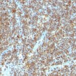
Variations in PCNA antibody staining intensity in immunohistochemistry on tissue sections are present across different anatomical locations. An intense signal was observed in glandular cells in the appendix, lymphoid tissue in appendix, hematopoietic cells in the bone marrow, glandular cells in the colon, germinal center cells in the lymph node, glandular cells in the small intestine and germinal center cells in the tonsil. More moderate antibody staining intensity was present in glandular cells in the appendix, lymphoid tissue in appendix, hematopoietic cells in the bone marrow, glandular cells in the colon, germinal center cells in the lymph node, glandular cells in the small intestine and germinal center cells in the tonsil. Low, but measureable presence of PCNA could be seen inglandular cells in the adrenal gland, cells in the granular layer in cerebellum, cells in the molecular layer in cerebellum, myocytes in heart muscle, macrophages in lung, pneumocytes in lung, respiratory epithelial cells in the nasopharynx, exocrine glandular cells in the pancreas, glandular cells in the parathyroid gland, trophoblastic cells in the placenta, myocytes in skeletal muscle, Langerhans in skin, smooth muscle cells in the smooth muscle, cells in the red pulp in spleen and glandular cells in the stomach. We were unable to detect PCNA in other tissues. Disease states, inflammation, and other physiological changes can have a substantial impact on antibody staining patterns. These measurements were all taken in tissues deemed normal or from patients without known disease.
Observed Antibody Staining Data By Tissue Type:
Tissues from cancer patients, for instance, have their own distinct pattern of PCNA expression as measured by anti-PCNA antibody immunohistochemical staining. The average level of expression by tumor is summarized in the table below. The variability row represents patient to patient variability in IHC staining.
| Sample Type | breast cancer | carcinoid | cervical cancer | colorectal cancer | endometrial cancer | glioma | head and neck cancer | liver cancer | lung cancer | lymphoma | melanoma | ovarian cancer | pancreatic cancer | prostate cancer | renal cancer | skin cancer | stomach cancer | testicular cancer | thyroid cancer | urothelial cancer |
|---|---|---|---|---|---|---|---|---|---|---|---|---|---|---|---|---|---|---|---|---|
| Signal Intensity | +++ | ++ | +++ | +++ | ++ | – | +++ | ++ | +++ | ++ | +++ | ++ | +++ | + | ++ | +++ | ++ | ++ | + | +++ |
| PCNA Variability | ++ | ++ | + | ++ | +++ | ++ | ++ | +++ | + | ++ | ++ | ++ | ++ | ++ | ++ | ++ | ++ | ++ | ++ | ++ |
| PCNA General Information | |
|---|---|
| Alternate Names | |
| Proliferating cell nuclear antigen, PCNA | |
| Molecular Weight | |
| 36kDa | |
| Chromosomal Location | |
| 20p12.3 | |
| Curated Database and Bioinformatic Data | |
| Gene Symbol | PCNA |
| Entrez Gene ID | 5111 |
| Ensemble Gene ID | ENSG00000132646 |
| RefSeq Protein Accession(s) | NP_872590, NP_002583 |
| RefSeq mRNA Accession(s) | NM_002592 NM_182649 |
| RefSeq Genomic Accession(s) | NC_000020, NG_047066, NC_018931 |
| UniProt ID(s) | P12004 |
| UniGene ID(s) | P12004 |
| HGNC ID(s) | 8729 |
| Cosmic ID(s) | PCNA |
| KEGG Gene ID(s) | hsa:5111 |
| PharmGKB ID(s) | PA263 |
| General Description of PCNA. | |
| Recognizes a non-histone protein of 36kDa, which is identified as proliferating cell nuclear antigen (PCNA). It is also known as cyclin or polymerase delta auxiliary protein. Elevated expression of PCNA/cyclin has been shown in the nucleus during late G1 phase immediately before the onset of DNA synthesis, becoming maximal during S-phase, declining during G2, M phases. This MAb is excellent for multiple applications. | |

There are no reviews yet.