
 PDF Datasheet
Human, Chimpanzee, Monkey, Goat, Rabbit, Rat, and Mouse Anti-Retinol Binding Protein-1 Antibody Product Attributes
Retinol Binding Protein-1 Previously Observed Antibody Staining Patterns
Observed Subcellular, Organelle Specific Staining Data:
Anti-RBP1 antibody staining is expected to be primarily localized to the cytosol and nucleoplasm.
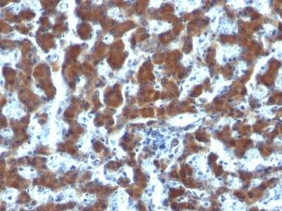
Observed Antibody Staining Data By Tissue Type:
Variations in Retinol Binding Protein-1 antibody staining intensity in immunohistochemistry on tissue sections are present across different anatomical locations. An intense signal was observed in Leydig cells in the testis. More moderate antibody staining intensity was present in Leydig cells in the testis. Low, but measureable presence of Retinol Binding Protein-1 could be seen inglandular cells in the breast, glial cells in the caudate nucleus, cells in the molecular layer in cerebellum, glandular cells in the cervix, uterine, cells in the endometrial stroma in endometrium, glandular cells in the gallbladder, glial cells in the hippocampus, cells in the tubules in kidney and cells in the seminiferous ducts in testis. We were unable to detect Retinol Binding Protein-1 in other tissues. Disease states, inflammation, and other physiological changes can have a substantial impact on antibody staining patterns. These measurements were all taken in tissues deemed normal or from patients without known disease.
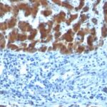
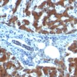
Observed Antibody Staining Data By Tissue Disease Status:
Tissues from cancer patients, for instance, have their own distinct pattern of Retinol Binding Protein-1 expression as measured by anti-Retinol Binding Protein-1 antibody immunohistochemical staining. The average level of expression by tumor is summarized in the table below. The variability row represents patient to patient variability in IHC staining.
| Sample Type | breast cancer | carcinoid | cervical cancer | colorectal cancer | endometrial cancer | glioma | head and neck cancer | liver cancer | lung cancer | lymphoma | melanoma | ovarian cancer | pancreatic cancer | prostate cancer | renal cancer | skin cancer | stomach cancer | testicular cancer | thyroid cancer | urothelial cancer |
|---|---|---|---|---|---|---|---|---|---|---|---|---|---|---|---|---|---|---|---|---|
| Signal Intensity | – | – | – | + | – | – | + | – | – | – | – | – | – | – | – | – | – | – | – | – |
| RBP1 Variability | + | ++ | + | ++ | ++ | ++ | ++ | + | ++ | + | + | ++ | ++ | + | + | ++ | + | ++ | + | ++ |
| Retinol Binding Protein-1 General Information | |
|---|---|
| Alternate Names | |
| Retinol binding protein 1, cellular RBP1, RBP1 | |
| Molecular Weight | |
| 21-25kDa | |
| Chromosomal Location | |
| 10q23.33 | |
| Curated Database and Bioinformatic Data | |
| Gene Symbol | RBP1 |
| Entrez Gene ID | 5947, 5948, 5950 |
| Ensemble Gene ID | ENSG00000114115, ENSG00000114113, ENSG00000138207 |
| RefSeq Protein Accession(s) | NP_001124464, NP_002890, NP_001124465, NP_004155, NP_001310446, NP_001310447, NP_006735 |
| RefSeq mRNA Accession(s) | NM_002899, NM_001130993, NM_001130992, NM_004164, NM_001323517, NM_001323518, NM_006744, |
| RefSeq Genomic Accession(s) | NC_018914, NG_047073, NC_000003, NC_000003, NC_018914NC_000010, NC_018921, NG_009104 |
| UniProt ID(s) | A0A0A0MQT0, P09455, P50120, Q5VY30, P02753 |
| UniGene ID(s) | A0A0A0MQT0, P09455, P50120, Q5VY30, P02753 |
| HGNC ID(s) | 9919, 9920, 9922 |
| Cosmic ID(s) | RBP1, RBP2, RBP4 |
| KEGG Gene ID(s) | hsa:5947, hsa:5948, hsa:5950 |
| PharmGKB ID(s) | PA34286, PA34287, PA34289 |
| General Description of Retinol Binding Protein-1. | |
| This MAb recognizes an epitope within the 74-182 C-terminal sequence (11kD peptide fragment) of human serum Cellular Retinol Binding Protein 1 (CRBP 1), a single-chain glycoprotein belonging to the superfamily of hydrophobic molecule transporter proteins, which is responsible for transport of retinol (vitamin A1) from the liver to peripheral target tissues, like the eye, where it mediates the cellular uptake. CRBP 1 is synthesized by hepatic parenchymal cells where it becomes bound to its ligand retinol, is then released into the circulation, where it binds further to the protein transthyretin, to form a transporting complex, which is big enough not to be lost by filtration through the kidney glomeruli. It is detected in nearly all tissues with higher expression in adult ovary, pancreas, pituitary gland, adrenal gland,, fetal liver. | |
There are no reviews yet.