
 PDF Datasheet
Human Anti-S100B Antibody Product Attributes
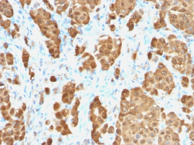
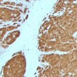
S100B Previously Observed Antibody Staining Patterns
Observed Subcellular, Organelle Specific Staining Data:
Anti-S100B antibody staining is expected to be primarily localized to the nucleus and vesicles.
Observed Antibody Staining Data By Tissue Type:
Variations in S100B antibody staining intensity in immunohistochemistry on tissue sections are present across different anatomical locations. An intense signal was observed in myoepithelial cells in the breast, glial cells in the caudate nucleus, neuronal cells in the caudate nucleus, cells in the granular layer in cerebellum, cells in the molecular layer in cerebellum, Purkinje cells in the cerebellum, glial cells in the cerebral cortex, neuronal cells in the cerebral cortex, neuropil in cerebral cortex, peripheral nerve/ganglion in colon, glial cells in the hippocampus, neuronal cells in the hippocampus, Langerhans in skin and peripheral nerve in mesenchymal tissue. More moderate antibody staining intensity was present in myoepithelial cells in the breast, glial cells in the caudate nucleus, neuronal cells in the caudate nucleus, cells in the granular layer in cerebellum, cells in the molecular layer in cerebellum, Purkinje cells in the cerebellum, glial cells in the cerebral cortex, neuronal cells in the cerebral cortex, neuropil in cerebral cortex, peripheral nerve/ganglion in colon, glial cells in the hippocampus, neuronal cells in the hippocampus, Langerhans in skin and peripheral nerve in mesenchymal tissue. Low, but measureable presence of S100B could be seen in. We were unable to detect S100B in other tissues. Disease states, inflammation, and other physiological changes can have a substantial impact on antibody staining patterns. These measurements were all taken in tissues deemed normal or from patients without known disease.
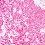
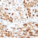
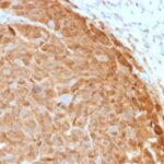
Observed Antibody Staining Data By Tissue Disease Status:
Tissues from cancer patients, for instance, have their own distinct pattern of S100B expression as measured by anti-S100B antibody immunohistochemical staining. The average level of expression by tumor is summarized in the table below. The variability row represents patient to patient variability in IHC staining.
| Sample Type | breast cancer | carcinoid | cervical cancer | colorectal cancer | endometrial cancer | glioma | head and neck cancer | liver cancer | lung cancer | lymphoma | melanoma | ovarian cancer | pancreatic cancer | prostate cancer | renal cancer | skin cancer | stomach cancer | testicular cancer | thyroid cancer | urothelial cancer |
|---|---|---|---|---|---|---|---|---|---|---|---|---|---|---|---|---|---|---|---|---|
| Signal Intensity | ++ | + | – | + | + | ++ | – | – | – | – | ++ | + | ++ | + | – | – | + | – | + | + |
| S100B Variability | ++ | ++ | ++ | ++ | ++ | ++ | ++ | ++ | + | + | +++ | ++ | + | ++ | + | + | ++ | ++ | ++ | ++ |
| S100B General Information | |
|---|---|
| Alternate Names | |
| S100 calcium-binding protein B, S100B | |
| Molecular Weight | |
| 10-12kDa | |
| Chromosomal Location | |
| 21q22.3 | |
| Curated Database and Bioinformatic Data | |
| Gene Symbol | S100B |
| Entrez Gene ID | 6285 |
| Ensemble Gene ID | ENSG00000160307 |
| RefSeq Protein Accession(s) | NP_006263, XP_016883913 |
| RefSeq mRNA Accession(s) | XM_017028424, NM_006272 |
| RefSeq Genomic Accession(s) | NC_018932, NC_000021 |
| UniProt ID(s) | A0A0S2Z4C5, P04271 |
| UniGene ID(s) | A0A0S2Z4C5, P04271 |
| HGNC ID(s) | 10500 |
| Cosmic ID(s) | S100B |
| KEGG Gene ID(s) | hsa:6285 |
| PharmGKB ID(s) | PA34912 |
| General Description of S100B. | |
| S100 belongs to the family of calcium binding proteins. S100A and S100B proteins are two members of the S100 family. S100A is composed of an alpha and a beta chain whereas S100B is composed of two beta chains. This antibody is specific against an epitope located on the beta-chain (i.e. in S-100A and S-100B) but not on the alpha-chain of S-100 (i.e. in S-100A and S100A0). This antibody can be used to localize S-100A and S-100B in various tissue sections. S-100 protein has been found in normal melanocytes, Langerhans cells, histiocytes, chondrocytes, lipocytes, skeletal and cardiac muscle, Schwann cells, epithelial and myoepithelial cells of the breast, salivary and sweat glands, as well as in glial cells. Neoplasms derived from these cells also express S-100 protein, albeit non-uniformly. A large number of well-differentiated tumors of the salivary gland, adipose and cartilaginous tissue, and Schwann cell-derived tumors express S-100 protein. Almost all malignant melanomas and cases of histiocytosis X are positive for S-100 protein. | |

-150x150.jpg)
There are no reviews yet.