

 PDF Datasheet
Human and Rat (-) Anti-Tenascin C Antibody Product Attributes
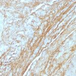
Tenascin C Previously Observed Antibody Staining Patterns
Observed Antibody Staining Data By Tissue Type:
Variations in Tenascin C antibody staining intensity in immunohistochemistry on tissue sections are present across different anatomical locations. Low, but measureable presence of Tenascin C could be seen inhematopoietic cells in the bone marrow, respiratory epithelial cells in the bronchus, squamous epithelial cells in the cervix, uterine, macrophages in lung, germinal center cells in the lymph node, non-germinal center cells in the lymph node, respiratory epithelial cells in the nasopharynx, squamous epithelial cells in the oral mucosa, ovarian stroma cells in the ovary, glandular cells in the prostate, rectum, salivary gland and seminal vesicle, myocytes in skeletal muscle, keratinocytes in skin, epidermal cells in the skin, cells in the red pulp in spleen, glandular cells in the stomach, germinal center cells in the tonsil and non-germinal center cells in the tonsil. We were unable to detect Tenascin C in other tissues. Disease states, inflammation, and other physiological changes can have a substantial impact on antibody staining patterns. These measurements were all taken in tissues deemed normal or from patients without known disease.
Observed Antibody Staining Data By Tissue Disease Status:
Tissues from cancer patients, for instance, have their own distinct pattern of Tenascin C expression as measured by anti-Tenascin C antibody immunohistochemical staining. The average level of expression by tumor is summarized in the table below. The variability row represents patient to patient variability in IHC staining.
| Sample Type | breast cancer | carcinoid | cervical cancer | colorectal cancer | endometrial cancer | glioma | head and neck cancer | liver cancer | lung cancer | lymphoma | melanoma | ovarian cancer | pancreatic cancer | prostate cancer | renal cancer | skin cancer | stomach cancer | testicular cancer | thyroid cancer | urothelial cancer |
|---|---|---|---|---|---|---|---|---|---|---|---|---|---|---|---|---|---|---|---|---|
| Signal Intensity | ++ | – | ++ | – | – | ++ | ++ | – | + | – | ++ | ++ | – | – | – | ++ | – | – | ++ | ++ |
| TNC Variability | ++ | ++ | ++ | ++ | ++ | + | + | ++ | ++ | ++ | ++ | ++ | ++ | ++ | ++ | ++ | ++ | ++ | ++ | ++ |
| Tenascin C General Information | |
|---|---|
| Alternate Names | |
| TNC, Tenascin C, TN-C | |
| Molecular Weight | |
| 210kDa, 300kDa | |
| Chromosomal Location | |
| 9q33 | |
| Curated Database and Bioinformatic Data | |
| Gene Symbol | TNC |
| Entrez Gene ID | 3371 |
| Ensemble Gene ID | ENSG00000041982 |
| RefSeq Protein Accession(s) | XP_016870167, XP_005252030, XP_006717164, XP_006717159, XP_005252029, XP_006717161, XP_005252031, XP_005252032, XP_011516928, XP_011516930, XP_016870168, XP_016870170, XP_006717160, XP_011516927, XP_011516931, XP_016870169, NP_002151 |
| RefSeq mRNA Accession(s) | XM_017014679, XM_017014681, XM_011518629, XM_011518626, XM_005251972, XM_006717097, XM_005251975, XM_011518625, XM_006717096, XM_011518628, XM_017014678, XM_017014680, NM_002160, XM_005251973, XM_006717098, XM_006717101 |
| RefSeq Genomic Accession(s) | NC_018920, NC_000009, NG_029637 |
| UniProt ID(s) | B4E1W8, P24821, Q4LE33, J3QSU6 |
| UniGene ID(s) | B4E1W8, P24821, Q4LE33, J3QSU6 |
| HGNC ID(s) | 5318 |
| Cosmic ID(s) | TNC |
| KEGG Gene ID(s) | hsa:3371 |
| PharmGKB ID(s) | PA35103 |
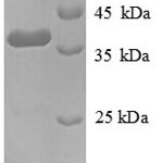
| General Description of Tenascin C. | |
| In Western Blot, it reacts with two bands of ~MW of 210kDa, 300kDa, identified as two isoforms of Tenascin C. Specificity of this MAb is validated by sequential immunoprecipitation with a PAb against Tenascin C. Tenascin C is a multifunctional, disulfide-linkedhexameric extracellular matrix glycoprotein expressed in association with mesenchymal epithelial interactions during development, in the neo-vasculature, stroma of undifferentiated tumors. In adults, it is restricted to certain epithelial-stromal interfaces, increases markedly in hyper-proliferative diseases, in stroma of many neoplasms, including gliomas, breast, squamous, lung carcinomas. | |
There are no reviews yet.