
 PDF Datasheet
Human Anti-Tyrosinase Antibody Product Attributes
Tyrosinase Previously Observed Antibody Staining Patterns
Observed Subcellular, Organelle Specific Staining Data:
Anti-TYR antibody staining is expected to be primarily localized to the vesicles.
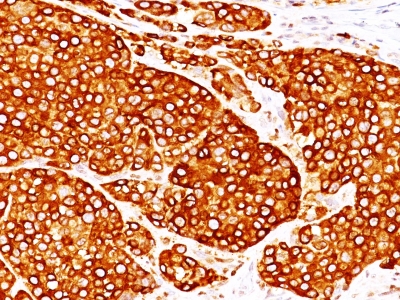
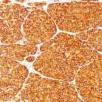
Observed Antibody Staining Data By Tissue Type:
Variations in Tyrosinase antibody staining intensity in immunohistochemistry on tissue sections are present across different anatomical locations. An intense signal was observed in melanocytes in skin. More moderate antibody staining intensity was present in melanocytes in skin. Low, but measureable presence of Tyrosinase could be seen in. We were unable to detect Tyrosinase in other tissues. Disease states, inflammation, and other physiological changes can have a substantial impact on antibody staining patterns. These measurements were all taken in tissues deemed normal or from patients without known disease.
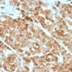
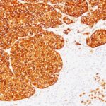
Observed Antibody Staining Data By Tissue Disease Status:
Tissues from cancer patients, for instance, have their own distinct pattern of Tyrosinase expression as measured by anti-Tyrosinase antibody immunohistochemical staining. The average level of expression by tumor is summarized in the table below. The variability row represents patient to patient variability in IHC staining.
| Sample Type | breast cancer | carcinoid | cervical cancer | colorectal cancer | endometrial cancer | glioma | head and neck cancer | liver cancer | lung cancer | lymphoma | melanoma | ovarian cancer | pancreatic cancer | prostate cancer | renal cancer | skin cancer | stomach cancer | testicular cancer | thyroid cancer | urothelial cancer |
|---|---|---|---|---|---|---|---|---|---|---|---|---|---|---|---|---|---|---|---|---|
| Signal Intensity | – | – | – | – | – | – | – | – | – | – | +++ | – | – | – | – | – | – | – | – | – |
| TYR Variability | + | + | + | + | + | + | + | + | + | + | + | + | + | + | + | + | + | + | + | + |
| Tyrosinase General Information | |
|---|---|
| Alternate Names | |
| Tyrosinase, Tumor Rejection Antigen AB | |
| Molecular Weight | |
| 70-80kDa | |
| Chromosomal Location | |
| 11q14.3 | |
| Curated Database and Bioinformatic Data | |
| Gene Symbol | TYR |
| Entrez Gene ID | 7299 |
| Ensemble Gene ID | ENSG00000077498 |
| RefSeq Protein Accession(s) | XP_011541272, NP_000363 |
| RefSeq mRNA Accession(s) | NM_000372, XM_011542970, |
| RefSeq Genomic Accession(s) | NC_000011, NG_008748, NC_018922 |
| UniProt ID(s) | P14679, L8B082 |
| UniGene ID(s) | P14679, L8B082 |
| HGNC ID(s) | 12442 |
| Cosmic ID(s) | TYR |
| KEGG Gene ID(s) | hsa:7299 |
| PharmGKB ID(s) | PA37095 |
| General Description of Tyrosinase. | |
| Recognizes a cluster of proteins between 70-80kDa, identified as tyrosinase. Occasionally a minor band at 55kDa is also detected. This MAb shows no cross-reaction with MAGE-1, tyrosinase-related protein 1, TRP-1/gp75. Tyrosinase is a copper-containing metalloglycoprotein that catalyzes several steps in the melanin pigment biosynthetic pathway; the hydroxylation of tyrosine to L-3,4-dihydroxy-phenylalanine (dopa),, the subsequent oxidation of dopa to dopaquinone. Mutations of the tyrosinase gene occur in various forms of albinism. Tyrosinase is one of the targets for cytotoxic T-cell recognition in melanoma patients. Staining of melanomas with this MAb shows tyrosinase in melanotic as well as amelanotic variants. This MAb is a useful marker for melanocytes, melanomas. | |
Reviews
There are no reviews yet.