
 PDF Datasheet
Human Anti-CD40 / TNFRSF5 Antibody Product Attributes
CD40 / TNFRSF5 / CD40L-Receptor Previously Observed Antibody Staining Patterns
Observed Antibody Staining Data By Tissue Type:
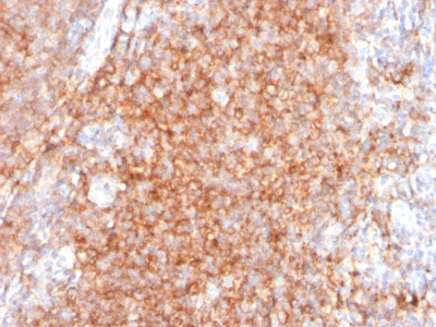
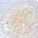
Variations in CD40 / TNFRSF5 antibody staining intensity in immunohistochemistry on tissue sections are present across different anatomical locations. An intense signal was observed in cells in the white pulp in spleen and germinal center cells in the lymph node and tonsil. More moderate antibody staining intensity was present in cells in the white pulp in spleen and germinal center cells in the lymph node and tonsil. Low, but measureable presence of CD40 / TNFRSF5 could be seen inlymphoid tissue in appendix and macrophages in lung. We were unable to detect CD40 / TNFRSF5 in other tissues. Disease states, inflammation, and other physiological changes can have a substantial impact on antibody staining patterns. These measurements were all taken in tissues deemed normal or from patients without known disease.
Observed Antibody Staining Data By Tissue Disease Status:
Tissues from cancer patients, for instance, have their own distinct pattern of CD40 / TNFRSF5 expression as measured by anti-CD40 / TNFRSF5 antibody immunohistochemical staining. The average level of expression by tumor is summarized in the table below. The variability row represents patient to patient variability in IHC staining.
| Sample Type | breast cancer | carcinoid | cervical cancer | colorectal cancer | endometrial cancer | glioma | head and neck cancer | liver cancer | lung cancer | lymphoma | melanoma | ovarian cancer | pancreatic cancer | prostate cancer | renal cancer | skin cancer | stomach cancer | testicular cancer | thyroid cancer | urothelial cancer |
|---|---|---|---|---|---|---|---|---|---|---|---|---|---|---|---|---|---|---|---|---|
| Signal Intensity | – | – | – | – | – | – | – | – | – | + | – | – | – | – | – | – | – | – | + | – |
| CD40 Variability | + | + | + | + | + | + | + | + | ++ | ++ | + | ++ | + | + | + | + | + | + | ++ | + |
| CD40 / TNFRSF5 / CD40L-Receptor General Information | |
|---|---|
| Alternate Names | |
| B-cell surface antigen CD40; Bp50; CD40; CD40L receptor; GP39; HIGM1; IGM; IMD3; p50; TBAM; TNF receptor superfamily member 5; TNFRSF5; TRAP | |
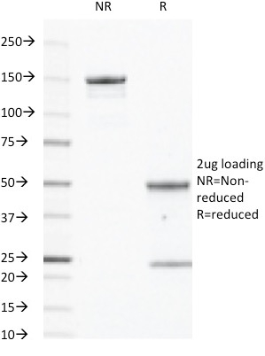
| Molecular Weight | |
| 43kDa | |
| Chromosomal Location | |
| Ships on blue ice. | |
| Curated Database and Bioinformatic Data | |
| Gene Symbol | 958 |
| Entrez Gene ID | CD40 |
| UniProt ID(s) | P25942 |
| UniGene ID(s) | Hs472860 |
| COSMIC ID Link(s) | CD40 |
| KEGG Gene ID(s) | hsa:958 |
| General Description of CD40 / TNFRSF5 / CD40L-Receptor. | |
| CD40 is a receptor on antigen-presenting cells of the immune system and is essential for mediating a broad variety of immune and inflammatory responses including T cell-dependent immunoglobulin class switching, memory B cell development, and germinal center formation. AT-hook transcription factor AKNA is reported to coordinately regulate the expression of this receptor and its ligand, which may be important for homotypic cell interactions. Adaptor protein TNFR2 interacts with this receptor and serves as a mediator of the signal transduction. The interaction of this receptor and its ligand is found to be necessary for amyloid-beta-induced microglial activation, and thus is thought to be an early event in Alzheimer disease pathogenesis. CD40 is expressed on B-lymphocytes, follicular dendritic cells, bone marrow-derived dendritic cells, thymic epithelium, and interdigitating cells in the T-cell zones of secondary lymphoid organs. | |

Reviews
There are no reviews yet.