
 PDF Datasheet
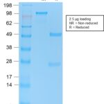
Human Anti-p53 Tumor Suppressor Protein Antibody Product Attributes
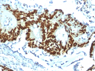
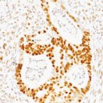
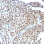
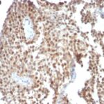
p53 Tumor Suppressor Protein Previously Observed Antibody Staining Patterns
Observed Subcellular, Organelle Specific Staining Data:
Variations in p53 Tumor Suppressor Protein antibody staining intensity in immunohistochemistry on tissue sections are present across different anatomical locations. Low, but measureable presence of p53 Tumor Suppressor Protein could be seen inglandular cells in the adrenal gland, hematopoietic cells in the bone marrow, glandular cells in the breast, respiratory epithelial cells in the bronchus, neuronal cells in the caudate nucleus, glandular cells in the cervix, uterine and endometrium, cells in the glomeruli in kidney, islets of Langerhans in pancreas, glandular cells in the parathyroid gland, decidual cells in the placenta, glandular cells in the salivary gland, fibroblasts in skin, cells in the white pulp in spleen, non-germinal center cells in the tonsil and squamous epithelial cells in the vagina. We were unable to detect p53 Tumor Suppressor Protein in other tissues. Disease states, inflammation, and other physiological changes can have a substantial impact on antibody staining patterns. These measurements were all taken in tissues deemed normal or from patients without known disease.
Observed Antibody Staining Data By Tissue Type:
Tissues from cancer patients, for instance, have their own distinct pattern of p53 Tumor Suppressor Protein expression as measured by anti-p53 Tumor Suppressor Protein antibody immunohistochemical staining. The average level of expression by tumor is summarized in the table below. The variability row represents patient to patient variability in IHC staining.
| Sample Type | breast cancer | carcinoid | cervical cancer | colorectal cancer | endometrial cancer | glioma | head and neck cancer | liver cancer | lung cancer | lymphoma | melanoma | ovarian cancer | pancreatic cancer | prostate cancer | renal cancer | skin cancer | stomach cancer | testicular cancer | thyroid cancer | urothelial cancer |
|---|---|---|---|---|---|---|---|---|---|---|---|---|---|---|---|---|---|---|---|---|
| Signal Intensity | – | – | + | + | – | – | – | – | + | – | + | – | ++ | – | – | + | + | + | – | ++ |
| TP53 Variability | + | + | ++ | ++ | + | ++ | ++ | ++ | ++ | ++ | +++ | ++ | ++ | + | + | ++ | ++ | ++ | + | +++ |
| p53 Tumor Suppressor Protein General Information | |
|---|---|
| Alternate Names | |
| Antigen NY-CO-13, BCC7, Cellular Tumor Antigen p53, LFS1, TP53, Transformation Related Protein 53 (TRP53), Tumor Protein p53, Tumor Suppressor p53 | |
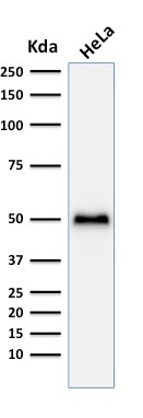
| Molecular Weight | |
| 53kDa | |
| Chromosomal Location | |
| Ships on blue ice. | |
| Curated Database and Bioinformatic Data | |
| Gene Symbol | 7157 |
| Entrez Gene ID | TP53 |
| UniProt ID(s) | P04637 |
| UniGene ID(s) | Hs654481 |
| COSMIC ID Link(s) | TP53 |
| KEGG Gene ID(s) | hsa:7157 |
| General Description of p53 Tumor Suppressor Protein. | |
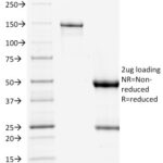
| The specificity of this monoclonal antibody to its intended target was validated by HuProtTM Array, containing more than 19,000, full-length human proteins. Recognizes a 53kDa protein, which is identified as p53 suppressor gene product. It reacts with the mutant as well as the wild form of p53 protein. p53 is a tumor suppressor gene expressed in a wide variety of tissue types and is involved in regulating cell growth, replication, and apoptosis. It binds to MDM2, SV40 T antigen and human papilloma virus E6 protein. Positive nuclear staining with p53 antibody has been reported to be a negative prognostic factor in breast carcinoma, lung carcinoma, colorectal, and urothelial carcinoma. Anti-p53 positivity has also been used to differentiate uterine serous carcinoma from endometrioid carcinoma as well as to detect intratubular germ cell neoplasia. Mutations involving p53 are found in a wide variety of malignant tumors, including breast, ovarian, bladder, colon, lung, and melanoma. | |
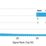
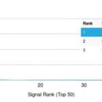
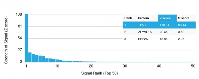
![Analysis of Mass Spec data (dashed-line) of fractions stained with p53 Tumor Suppressor Protein <a href="https://enquirebio.com/validation-project-details/" target="_blank">MS-QAVA™ monoclonal antibody</a> [Clone: BP53-12] (solid-line), reveals that less than 12.9% of signal is attributable to non-specific binding of anti-p53 Tumor Suppressor Protein [Clone BP53-12] to targets other than TP53 protein. Even frequently cited antibodies have much greater non-specific interactions, averaging over 30%. Data in image is from analysis in A431, RT4 and MCF7 cells.](https://cdn-enquirebio.pressidium.com/wp-content/uploads/2017/10/enQuire-Bio-7157-MSM1-P1-anti-p53-Tumor-Suppressor-Protein-antibody-150x150.png)
-150x150.jpg)
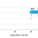
![Analysis of Mass Spec data (dashed-line) of fractions stained with p53 Tumor Suppressor Protein <a href="https://enquirebio.com/validation-project-details/" target="_blank">MS-QAVA™ monoclonal antibody</a> [Clone: BP53-12 + DO-7] (solid-line), reveals that less than 10.8% of signal is attributable to non-specific binding of anti-p53 Tumor Suppressor Protein [Clone BP53-12 + DO-7] to targets other than TP53 protein. Even frequently cited antibodies have much greater non-specific interactions, averaging over 30%. Data in image is from analysis in A431, RT4 and MCF7 cells.](https://cdn-enquirebio.pressidium.com/wp-content/uploads/2017/10/enQuire-Bio-7157-MSM3-P1-anti-p53-Tumor-Suppressor-Protein-antibody-150x150.png)
![Analysis of Mass Spec data (dashed-line) of fractions stained with p53 Tumor Suppressor Protein <a href="https://enquirebio.com/validation-project-details/" target="_blank">MS-QAVA™ monoclonal antibody</a> [Clone: DO-7] (solid-line), reveals that less than 12.1% of signal is attributable to non-specific binding of anti-p53 Tumor Suppressor Protein [Clone DO-7] to targets other than TP53 protein. Even frequently cited antibodies have much greater non-specific interactions, averaging over 30%. Data in image is from analysis in A431, RT4 and MCF7 cells.](https://cdn-enquirebio.pressidium.com/wp-content/uploads/2017/10/enQuire-Bio-7157-MSM2-P1-anti-p53-Tumor-Suppressor-Protein-antibody-150x150.png)
There are no reviews yet.