
%20prostate%20adenocarcinoma.jpg)
 PDF Datasheet
Human and Mouse (-) Anti-Androgen Receptor Antibody Product Attributes
Androgen Receptor Previously Observed Antibody Staining Patterns
Observed Subcellular, Organelle Specific Staining Data:
Anti-ar antibody staining is expected to be primarily localized to the mitochondria.
Observed Antibody Staining Data By Tissue Type:
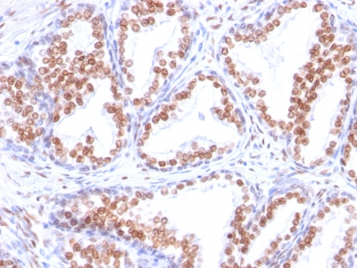
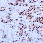
Variations in Androgen Receptor antibody staining intensity in immunohistochemistry on tissue sections are present across different anatomical locations. An intense signal was observed in glandular cells in the epididymis, seminal vesicle. More moderate antibody staining intensity was present in glandular cells in the epididymis, seminal vesicle. Low, but measureable presence of Androgen Receptor could be seen inglandular cells in the cervix, uterine. We were unable to detect Androgen Receptor in other tissues. Disease states, inflammation, and other physiological changes can have a substantial impact on antibody staining patterns. These measurements were all taken in tissues deemed normal or from patients without known disease.
Observed Antibody Staining Data By Tissue Disease Status:
Tissues from cancer patients, for instance, have their own distinct pattern of Androgen Receptor expression as measured by anti-Androgen Receptor antibody immunohistochemical staining. The average level of expression by tumor is summarized in the table below. The variability row represents patient to patient variability in IHC staining.
| Sample Type | breast cancer | carcinoid | cervical cancer | colorectal cancer | endometrial cancer | glioma | head and neck cancer | liver cancer | lung cancer | lymphoma | melanoma | ovarian cancer | pancreatic cancer | prostate cancer | renal cancer | skin cancer | stomach cancer | testicular cancer | thyroid cancer | urothelial cancer |
|---|---|---|---|---|---|---|---|---|---|---|---|---|---|---|---|---|---|---|---|---|
| Signal Intensity | ++ | – | – | – | – | – | – | – | – | – | – | – | – | +++ | – | – | – | – | – | – |
| AR Variability | ++ | + | + | + | ++ | + | ++ | + | + | + | + | ++ | + | + | + | ++ | + | + | + | + |
| Androgen Receptor General Information | |
|---|---|
| Alternate Names | |
| AR, Androgen Receptor, NR3C4 | |
| Molecular Weight | |
| 110kDa | |
| Chromosomal Location | |
| Xq12 | |
| Curated Database and Bioinformatic Data | |
| Gene Symbol | AR |
| Entrez Gene ID | 367 |
| Ensemble Gene ID | ENSG00000169083 |
| RefSeq Protein Accession(s) | NP_000035, NP_001334992, NP_001334993, NP_001011645, NP_001334990 |
| RefSeq mRNA Accession(s) | NM_001348063, NM_001348064 NM_001011645, NM_001348061, NM_000044 |
| RefSeq Genomic Accession(s) | NC_018934, NG_009014, NC_000023 |
| UniProt ID(s) | F1D8N5, A0A087WUX9, G4VV16, Q9NUA2, P10275 |
| UniGene ID(s) | F1D8N5, A0A087WUX9, G4VV16, Q9NUA2, P10275 |
| HGNC ID(s) | 644 |
| Cosmic ID(s) | AR |
| KEGG Gene ID(s) | hsa:367 |
| PharmGKB ID(s) | PA57 |
| General Description of Androgen Receptor. | |
| Recognizes a protein of 110kDa, which is identified as,rogen receptor (AR). It reacts with full length,, the newly described A form of the receptor. It does not cross react with estrogen, progesterone, or glucocorticoid receptors. The expression of AR is reportedly inversely correlated with histologic grade i.e. well differentiated prostate tumors show higher expression than the poorly differentiated tumors. In prostate cancer, AR has been proposed, as a marker of hormone-responsiveness, thus it may be useful in identifying patients likely to benefit from anti-androgen therapy. Anti-androgen receptor has been useful clinically in differentiating morpheaform basal cell carcinoma (mBCC) from desmoplastic trichoepithelioma (DTE) in the skin.This MAb is superb for staining of formalin/paraffin tissues. | |
-150x150.jpg)
-150x150.jpg)
-150x150.jpg)
Reviews
There are no reviews yet.