
Human Anti-CD45 / LCA Antibody Product Attributes
Species: Human
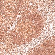
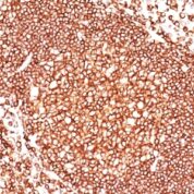
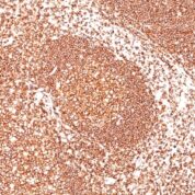
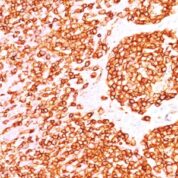
Tested Applications: Flow Cytometry, Immunofluorescence, Immunohistochemistry (IHC).
Application Notes: Flow Cytometry (0.5-1ug of antibody/million cells in 0.1ml), Immunofluorescence (0.5-1ug of antibody/ml), Immunohistochemistry (IHC) (Formalin-fixed) (0.5-1.0ug of antibody/ml for 30 minutes at RT)
Clonality: Monoclonal
Anti-CD45 / LCA Antibody Clone: 135-4C5
Clone 135-4C5 Host and Isotype: Mouse IgG2b kappa
Anti-Human CD45 / LCA Positive Control Sample: Ramos, U-698, or GA-10 cells. Tonsil
Cellular Localization of Antibody <135-4C5 Staining: Cell surface, cytoplasmic
Buffer and Stabilizer: 10mM PBS with 0.05% BSA & 0.05% azide.
Antibody Concentration: 200ug/ml
Antibody Purification Method:Protein A/G Purified
Immunogen: Stimulated human leukocytes
Storage Conditions: Store at 2 to 8° C (refrigerate). Stable for 24 months when properly stored.
CD45 / LCA Previously Observed Antibody Staining Patterns
Observed Antibody Staining Data By Tissue Type:
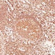
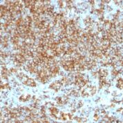
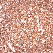
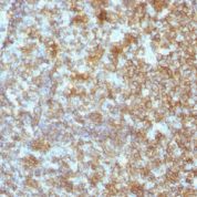
Variations in CD45 / LCA antibody staining intensity in immunohistochemistry on tissue sections are present across different anatomical locations. An intense signal was observed in lymphoid tissue in appendix, hematopoietic cells in the bone marrow, germinal center cells in the lymph node, non-germinal center cells in the lymph node, cells in the red pulp in spleen, cells in the white pulp in spleen, germinal center cells in the tonsil and non-germinal center cells in the tonsil. More moderate antibody staining intensity was present in lymphoid tissue in appendix, hematopoietic cells in the bone marrow, germinal center cells in the lymph node, non-germinal center cells in the lymph node, cells in the red pulp in spleen, cells in the white pulp in spleen, germinal center cells in the tonsil and non-germinal center cells in the tonsil. Low, but measureable presence of CD45 / LCA could be seen in. We were unable to detect CD45 / LCA in other tissues. Disease states, inflammation, and other physiological changes can have a substantial impact on antibody staining patterns. These measurements were all taken in tissues deemed normal or from patients without known disease.Observed Antibody Staining Data By Tissue Disease Status:
Tissues from cancer patients, for instance, have their own distinct pattern of CD45 / LCA expression as measured by anti-CD45 / LCA antibody immunohistochemical staining. The average level of expression by tumor is summarized in the table below. The variability row represents patient to patient variability in IHC staining.| Sample Type | breast cancer | carcinoid | cervical cancer | colorectal cancer | endometrial cancer | glioma | head and neck cancer | liver cancer | lung cancer | lymphoma | melanoma | ovarian cancer | pancreatic cancer | prostate cancer | renal cancer | skin cancer | stomach cancer | testicular cancer | thyroid cancer | urothelial cancer |
|---|---|---|---|---|---|---|---|---|---|---|---|---|---|---|---|---|---|---|---|---|
| Signal Intensity | - | - | - | - | - | - | - | - | - | +++ | - | - | - | - | - | - | - | - | - | - |
| PTPRC Variability | + | + | + | + | + | + | + | + | + | + | + | + | + | + | + | + | + | + | + | + |
Limitations and Warranty
enQuire Bio's CD45 / LCA Anti-Human Monoclonal is available for Research Use Only. This antibody is guaranteed to work for a period of two years when properly stored.

Zoey Niu
I have tried a ton of CD45 primary antibodies and finally decided this one. For me, it recognizes CD45 on leucocytes in the bloodstream. The picture shows a spike experiment (cancer cell into healthy blood), CK in Cy3, CD45 shows up in green. I have two other isotypes in the panel for multi-color staining: mouse IgG2a and mouse IgG1. They do not cross-react if use the correct secondary.