


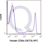
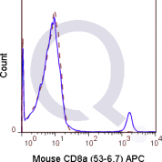
Human Anti-CD8 Antibody Product Attributes
CD8 Previously Observed Antibody Staining Patterns
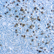
Observed Antibody Staining Data By Tissue Type:
Variations in CD8 antibody staining intensity in immunohistochemistry on tissue sections are present across different anatomical locations. An intense signal was observed in cells in the red pulp in spleen, cells in the white pulp in spleen, lymphoid tissue in appendix and non-germinal center cells in the lymph node and tonsil. More moderate antibody staining intensity was present in cells in the red pulp in spleen, cells in the white pulp in spleen, lymphoid tissue in appendix and non-germinal center cells in the lymph node and tonsil. Low, but measureable presence of CD8 could be seen inmacrophages in lung. We were unable to detect CD8 in other tissues. Disease states, inflammation, and other physiological changes can have a substantial impact on antibody staining patterns. These measurements were all taken in tissues deemed normal or from patients without known disease.
| CD8 General Information | |
|---|---|
| Alternate Names | |
| CD8A, MAL, CD8, p32, Leu2, MAL | |
| Curated Database and Bioinformatic Data | |
| Gene Symbol | CD8A |
| Entrez Gene ID | 925 |
| Ensemble Gene ID | ENSG00000153563 |
| RefSeq Protein Accession(s) | NP_001759, NP_001139345, NP_741969 |
| RefSeq mRNA Accession(s) | NM_001145873, NM_001768, NM_171827, NR_027353, |
| RefSeq Genomic Accession(s) | NC_000002, NG_011608, NC_018913 |
| UniProt ID(s) | P01732, Q8TAW8, Q6ZVS2 |
| UniGene ID(s) | P01732, Q8TAW8, Q6ZVS2 |
| HGNC ID(s) | 1706 |
| Cosmic ID(s) | CD8A |
| KEGG Gene ID(s) | hsa:925 |
| PharmGKB ID(s) | PA26244 |
| General Description of CD8. | |
| The RPA-T8 antibody is specific for the 32-34 kDa alpha chain of human CD8, known as CD8a or CD8 alpha. CD8a can form a homodimer (CD8 alpha-alpha), but is more commonly expressed as a heterodimer with a second chain known as CD8b or CD8 beta. CD8 acts as a co-receptor for antigen recognition and subsequent T cell activation that is initiated upon binding of the T cell receptor (TCR) to antigen-bearing MHC Class I molecules. The cytoplasmic domains of CD8 provide binding sites for the tyrosine kinase lck, facilitating intracellular signaling events that lead to T cell activation, development, and cytotoxic effector functions. CD8+ cytotoxic T cells (CTLs) play an important role in inducing cell death of tumor cells, as well as cells infected by virus, bacteria or parasites.The RPA-T8 antibody is widely used as a phenotypic marker for CD8 on cytotoxic T cells, thymocytes, as well as on certain cell types that do not also express the TCR, including some NK cells and lymphoid dendritic cells. It is cross-reactive with CD8 in several non-human species, including Baboon, Chimpanzee, Cynomolgus and Rhesus. If used together with an alternative Human Anti-CD8a clone, Hit8a, the RPA-T8 antibody will not block binding of Hit8a to CD8a. | |

There are no reviews yet.