Immunohistochemical staining of EpCAM of human FFPE tissue followed by incubation with HRP labeled secondary and development with DAB substrate.

EpCAM [E29] Antibody (cGMP).

$ 135.00$ 395.00
Please Select Product Options Below To View The Catalog Number.

SKU: QIVD58
Species: Human
Tested Applications: IHC [IVD]
Available Conjugates:
Isotype: Mouse IgG2a, kappa

Product NumberDescriptionPrice
QIVD58-0.1ml Size: 0.1 ml, Format: Concentrate $ 135.00
QIVD58-0.5ml Size: 0.5 ml, Format: Concentrate $ 235.00
QIVD58-1ml Size: 1 ml, Format: Concentrate $ 395.00
Shipping Information
In Stock
Flat Rate Shipping Anywhere in the US: $45
Datasheets and Documentation
Product Datasheet
Certificate of Analysis and Tags (Coming Soon)
Lot Number:

Expiration Date:

Concentration (Write Lyophilized if Lyophilized):

Reconsitution Instructions (Leave Blank if Liquid):

Manufacture Date:

Purity:

Bioactivity (test results eg. IU/ml):


SKU: QIVD58-0.1ml
EpCAM General Information
Alternate Names
Molecular Weight
34.9 kDa
Chromosomal Location
p21 [chr: 2] [chr_start: 47345158] [chr_end: 47387601] [strand: 1]
Curated Database and Bioinformatic Data
Gene SymbolEPCAM
Entrez Gene ID4072
RefSeq Protein Accession(s)NP_002345
RefSeq mRNA Accession(s)NM_002354;
RefSeq Genomic Accession(s)NG_012352; NC_000002
UniProt ID(s)P16422
PharmGKB ID(s)PA35493
KEGG Gene ID(s)hsa:4072
Associated Diseases (KEGG IDs)Diarrhea 5, with tufting enteropathy, congenital (DIAR5) [MIM:613217]: An intractable diarrhea of infancy characterized by villous atrophy and absence of inflammation, with intestinal epithelial cell dysplasia manifesting as focal epithelial tufts in the duodenum and jejunum. {ECO:0000269|PubMed:18572020}. The disease is caused by mutations affecting the gene represented in this entry.; Hereditary non-polyposis colorectal cancer 8 (HNPCC8) [MIM:613244]: An autosomal dominant disease associated with marked increase in cancer susceptibility. It is characterized by a familial predisposition to early-onset colorectal carcinoma (CRC) and extra-colonic tumors of the gastrointestinal, urological and female reproductive tracts. HNPCC is reported to be the most common form of inherited colorectal cancer in the Western world. Clinically, HNPCC is often divided into two subgroups. Type I is characterized by hereditary predisposition to colorectal cancer, a young age of onset, and carcinoma observed in the proximal colon. Type II is characterized by increased risk for cancers in certain tissues such as the uterus, ovary, breast, stomach, small intestine, skin, and larynx in addition to the colon. Diagnosis of classical HNPCC is based on the Amsterdam criteria: 3 or more relatives affected by colorectal cancer, one a first degree relative of the other two; 2 or more generation affected; 1 or more colorectal cancers presenting before 50 years of age; exclusion of hereditary polyposis syndromes. The term 'suspected HNPCC' or 'incomplete HNPCC' can be used to describe families who do not or only partially fulfill the Amsterdam criteria, but in whom a genetic basis for colon cancer is strongly suspected. {ECO:0000269|PubMed:19098912}. The disease is caused by mutations affecting the gene represented in this entry. HNPCC8 results from heterozygous deletion of 3-prime exons of EPCAM and intergenic regions directly upstream of MSH2, resulting in transcriptional read-through and epigenetic silencing of MSH2 in tissues expressing EPCAM.
General Description of EpCAM.
This antibody reacts with an antigen of 265-400 kDa identified as epithelial membrane antigen (EMA) or MUC-1. EMA belonging to a heterogeneous group of heavily glycosylated proteins called human milk fat globule proteins. It stains both normal and neoplastic cells. Among normal epithelia, it reacts strongly with mammary epithelium and glandular epithelia but shows a patchy staining with squamous epithelium.

Antibody (Suitable for clinical applications)

Sample Type: FFPE Patient Samples.
Tested Applications: IHC. Approved for In Vitro Diagnostic Procedures on FFPE tissues. For tissue collection recommendations, please see datasheet sent with product.
Application Notes
Specification Recommendation
Recommended Dilution (Conc) 1:50-1:100
Pretreatment Citrate Buffer pH 6.0
Incubation Parameters 30 min at Room Temperature

Prior to use, inspect vial for the presence of any precipitate or other unusual physical properties. These can indicate that the antibody has degraded and is no longer suitable for patient samples. Please run positive and negative controls simultaneously with all patient samples to account and control for errors in laboratory procedure. Use of methods or materials not recommended by enQuire Bio including change to dilution range and detection system should be routinely validated by the user.

Clonality: Monoclonal
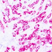
Anti-EpCAM Antibody Clone: E29
Host and Isotype: Mouse IgG2a, kappa
Recommended Positive Control Sample: Breast carcinoma
Cellular Localization of Antibody E29 Staining: Cytoplasmic, cell membrane
Buffer and Stabilizer: PBS with 1% BSA and 0.05% NaN3
Antibody Concentration: Lot specific. Plese contact tech support for data.
Immunogen: BALB/C mice were immunized with delipidated human milk fat globule membrane preparation.
Storage Conditions: This antibody should be stored refrigerated (2-8°C). This product should not be used past the expiration date printed on the vial.

EpCAM Information for Pathologists

Summary:

Antibody to cell membrane glycoproteins expressed on healthy epithelia and in various carcinomas. Also known as epithelial cell adhesion molecule, BerEp4 (Ber-EP4, J Clin Pathol 1990;43:213), MOC31 (MOC-31, Acta Neuropathol 1991;83:46), CD326, TACSTD1 protein. MOC31 and BerEP4 are both anti-EpCAM antibodies, MOC31 appears to be superior (Appl Immunohistochem Mol Morphol 2009;17:202). Anti-EpCam antibodies are in clinical trials for patients with cancer (Br J Cancer 2007;96:417). Uses by pathologists

Common Uses By Pathologists:

Membranous staining. Sensitive and specific for lung adenocarcinoma (positive) vs. mesothelioma (negative, Am J Surg Pathol 2001;25:43). May help distinguish, as part of a panel, hepatocellular carcinoma (usually negative) from metastatic adenocarcinoma to liver or cholangiocarcinoma (usually positive, Mod Pathol 2002;15:1279). Immunoexpression may predict poor survival in carcinomas of breast, gallbladder (Am J Clin Pathol 2008;129:424), ovary, pancreas. Microscopic (histologic) images

Limitations and Warranty

This antibody is manufactured in accordance with clinical good manufacturing practices in an ISO13485:2016 certified production facility. It is intended for multiple uses including in vitro diagnostic use and research use only applications. Please see vial label for expiration date. We strive to always deliver antibodies with a shelf life of at least two years.

There are no reviews yet.

Be the first to review “EpCAM [E29] Antibody (cGMP).”

Your email address will not be published. Required fields are marked *

Share a Protocol (View Reviewed Protocols Below)

Protocol Title

Materials

Methods

Notes


  protocol submission gif
enQuire Bio, LLC   8420 S Continental Divide Rd, #202   Littleton, CO 80127
Chat: See lower right corner. | enquire@enquirebio.com | Fax: 1-720-897-3730
Customer Service Available M-F 8AM-6PM MST or 24/7 Via Email

EpCAM [E29] Antibody (cGMP).