

Human specific, Mouse (-), and Rat (-) Anti-HSP60 Antibody Product Attributes
HSP60 Previously Observed Antibody Staining Patterns
Observed Subcellular, Organelle Specific Staining Data:
Anti-HSPD1 antibody staining is expected to be primarily localized to the mitochondria. There is variability in either the signal strength or the localization of signal in mitochondria from cell to cell.
Observed Antibody Staining Data By Tissue Type:
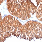
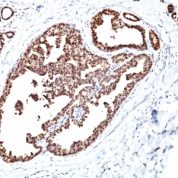
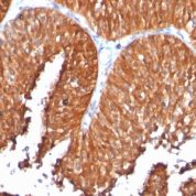
Variations in HSP60 antibody staining intensity in immunohistochemistry on tissue sections are present across different anatomical locations. An intense signal was observed in cells in the tubules in kidney, glandular cells in the adrenal gland, duodenum, fallopian tube and rectum, hepatocytes in liver and respiratory epithelial cells in the bronchus. More moderate antibody staining intensity was present in cells in the tubules in kidney, glandular cells in the adrenal gland, duodenum, fallopian tube and rectum, hepatocytes in liver and respiratory epithelial cells in the bronchus. Low, but measureable presence of HSP60 could be seen inadipocytes in mesenchymal tissue, cells in the red pulp in spleen, cells in the seminiferous ducts in testis, cells in the white pulp in spleen, decidual cells in the placenta, fibroblasts in mesenchymal tissue, glandular cells in the breast, cervix, uterine and thyroid gland, glial cells in the caudate nucleus, cerebral cortex and hippocampus, islets of Langerhans in pancreas, Leydig cells in the testis, macrophages in lung, melanocytes in skin, myocytes in heart muscle, neuronal cells in the caudate nucleus, cerebral cortex and hippocampus, non-germinal center cells in the lymph node and tonsil, ovarian stroma cells in the ovary, peripheral nerve in mesenchymal tissue, Purkinje cells in the cerebellum, squamous epithelial cells in the oral mucosa and urothelial cells in the urinary bladder. We were unable to detect HSP60 in other tissues. Disease states, inflammation, and other physiological changes can have a substantial impact on antibody staining patterns. These measurements were all taken in tissues deemed normal or from patients without known disease.
Observed Antibody Staining Data By Tissue Disease Status:
Tissues from cancer patients, for instance, have their own distinct pattern of HSP60 expression as measured by anti-HSP60 antibody immunohistochemical staining. The average level of expression by tumor is summarized in the table below. The variability row represents patient to patient variability in IHC staining.
| Sample Type | breast cancer | carcinoid | cervical cancer | colorectal cancer | endometrial cancer | glioma | head and neck cancer | liver cancer | lung cancer | lymphoma | melanoma | ovarian cancer | pancreatic cancer | prostate cancer | renal cancer | skin cancer | stomach cancer | testicular cancer | thyroid cancer | urothelial cancer |
|---|---|---|---|---|---|---|---|---|---|---|---|---|---|---|---|---|---|---|---|---|
| Signal Intensity | +++ | +++ | ++ | +++ | +++ | ++ | +++ | +++ | +++ | ++ | +++ | ++ | +++ | +++ | +++ | ++ | +++ | +++ | +++ | +++ |
| HSPD1 Variability | + | + | ++ | + | + | ++ | + | + | + | ++ | ++ | ++ | ++ | + | + | ++ | ++ | + | ++ | ++ |
| HSP60 General Information | |
|---|---|
| Alternate Names | |
| GroEL | |
| Molecular Weight | |
| 60kDa | |
| Chromosomal Location | |
| 2q33.1 | |
| Curated Database and Bioinformatic Data | |
| Gene Symbol | HSPD1 |
| Entrez Gene ID | 3329 |
| Ensemble Gene ID | ENSG00000144381 |
| RefSeq Protein Accession(s) | NP_002147, NP_955472 |
| RefSeq mRNA Accession(s) | NM_199440 NM_002156 |
| RefSeq Genomic Accession(s) | NC_000002, NC_018913, NG_008915 |
| UniProt ID(s) | P10809, A0A024R3X4 |
| UniGene ID(s) | P10809, A0A024R3X4 |
| HGNC ID(s) | 5261 |
| Cosmic ID(s) | HSPD1 |
| KEGG Gene ID(s) | hsa:3329 |
| PharmGKB ID(s) | PA29527 |
| General Description of HSP60. | |
| Recognizes a 60kDa protein, identified as the heat shock protein 60 (hsp60). A wide variety of environmental, pathophysiological stressful conditions trigger the synthesis of a family of proteins known as heat shock proteins (hsps), more appropriately called as stress response proteins (srps). Hsp60 is a potential antigen in a number of autoimmune diseases. In human arthritis, in experimentally induced arthritis in animals, disease development coincides with the development of immune reactivity directed against not only bacterial hsp60, but also against its mammalian homolog. Clone GROEL730 reacts only with human, is useful in distinguishing human hsp60 from other mammalian, bacterial hsp60. | |

There are no reviews yet.