

Antibody (Suitable for clinical applications)
Sample Type: FFPE Patient Samples.
Tested Applications: IHC. Approved for In Vitro Diagnostic Procedures on FFPE tissues. For tissue collection recommendations, please see datasheet sent with product.
Application Notes
| Specification | Recommendation |
|---|---|
| Recommended Dilution (Conc) | 1:50-1:100 |
| Pretreatment | EDTA Buffer pH 8.0 |
| Incubation Parameters | 30 min at Room Temperature |
Prior to use, inspect vial for the presence of any precipitate or other unusual physical properties. These can indicate that the antibody has degraded and is no longer suitable for patient samples. Please run positive and negative controls simultaneously with all patient samples to account and control for errors in laboratory procedure. Use of methods or materials not recommended by enQuire Bio including change to dilution range and detection system should be routinely validated by the user.
Clonality: Monoclonal
Anti-PD1 Antibody Clone: DBM15.5
Host and Isotype: Mouse IgG1, kappa
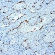
Recommended Positive Control Sample: Squamous lung carcinoma
Cellular Localization of Antibody DBM15.5 Staining: Cytoplasm and cell surface
Buffer and Stabilizer: PBS with 1% BSA and 0.05% NaN3
Antibody Concentration: Lot specific. Plese contact tech support for data.
Immunogen: Recombinant full-length human PDCD1 protein.
Storage Conditions: This antibody should be stored refrigerated (2-8°C). This product should not be used past the expiration date printed on the vial.
PD1 Information for Pathologists
Summary:
Also known as CD279. Coinhibitory receptor of lymphocytes / other immune system cells. Controls lymphocyte activation by providing negative signals in conjunction with signals from lymphocyte antigen receptors (Future Oncol 2011;7:929). Expressed by germinal center associated helper T cells; inhibits T cell activity (Hum Pathol 2009;40:1715). Expressed by CD8+ T cells, associated with CD8 activation (AIDS Res Hum Retroviruses 2012;28:465).Common Uses By Pathologists:
Differentiate primary cutaneous CD4 small / medium sized pleomorphic T cell lymphoma and cutaneous pseudo-T cell lymphomas (PD-1 positive) from other cutaneous T cell lymphomas (usually PD-1 negative, Am J Surg Pathol 2012;36:109). PD-1+ rosettes of T cells around neoplastic cells is relatively specific for nodular lymphocyte predominant Hodgkin lymphoma (Hum Pathol 2009;40:1715). Microscopic (histologic) images Images hosted on PathOut server:. Contributed by GenomeMe:.Limitations and Warranty
This antibody is manufactured in accordance with clinical good manufacturing practices in an ISO13485:2016 certified production facility. It is intended for multiple uses including in vitro diagnostic use and research use only applications. Please see vial label for expiration date. We strive to always deliver antibodies with a shelf life of at least two years.

There are no reviews yet.