Analysis of Protein Array containing more than 19,000 full-length human proteins using Filaggrin Mouse Monoclonal Antibody (FLG/1945). Z- and S- Score: The Z-score represents the strength of a signal that a monoclonal antibody (MAb) (in combination with a fluorescently-tagged anti-IgG secondary antibody) produces when binding to a particular protein on the HuProtTM array. Z-scores are described in units of standard deviations (SD’s) above the mean value of all signals generated on that array. If targets on HuProtTM are arranged in descending order of the Z-score, the S-score is the difference (also in units of SD’s) between the Z-score. S-score therefore represents the relative target specificity of a MAb to its intended target.  A MAb is considered to specific to its intended target, if the MAb has an S-score of at least 2.5. For example, if a MAb binds to protein X with a Z-score of 43 and to protein Y with a Z-score of 14, then the S-score for the binding of that MAb to protein X is equal to 29.
Analysis of Protein Array containing more than 19,000 full-length human proteins using Filaggrin Mouse Monoclonal Antibody (FLG/1945). Z- and S- Score: The Z-score represents the strength of a signal that a monoclonal antibody (MAb) (in combination with a fluorescently-tagged anti-IgG secondary antibody) produces when binding to a particular protein on the HuProtTM array. Z-scores are described in units of standard deviations (SD’s) above the mean value of all signals generated on that array. If targets on HuProtTM are arranged in descending order of the Z-score, the S-score is the difference (also in units of SD’s) between the Z-score. S-score therefore represents the relative target specificity of a MAb to its intended target.  A MAb is considered to specific to its intended target, if the MAb has an S-score of at least 2.5. For example, if a MAb binds to protein X with a Z-score of 43 and to protein Y with a Z-score of 14, then the S-score for the binding of that MAb to protein X is equal to 29.SDS-PAGE Analysis Purified Filaggrin Mouse Monoclonal Antibody (FLG/1945).Confirmation of Integrity and Purity of Antibody.

Filaggrin (Keratinocyte Differentiation Marker) Ultraspecific Antibody

Tested against >20,000 Human Proteins

$ 228.00$ 488.00
Please Select Product Options Below To View The Catalog Number.

SKU: 2312-MSM5
Species: Human
Tested Applications: IHC
Available Conjugates: Unconjugated
Isotype: Mouse IgG2b, kappa
Mass Spec Validated?: Not MS Validated

Product NumberDescriptionPrice
2312-MSM5-P0 Size: 20 ug, Tag: Unconjugated $ 199.00
2312-MSM5-P1 Size: 100 ug, Tag: Unconjugated $ 429.00
2312-MSM5-P1ABX Size: 100 ug, Tag: Unconjugated $ 429.00
Shipping Information
In Stock.
Flat Rate Shipping Anywhere in the US: $45
Datasheets and Documentation
Product Datasheet
Certificate of Analysis and Tags (Coming Soon)
Lot Number:

Expiration Date:

Concentration (Write Lyophilized if Lyophilized):

Reconsitution Instructions (Leave Blank if Liquid):

Manufacture Date:

Purity:

Bioactivity (test results eg. IU/ml):


SKU: 2312-MSM5-P0
Filaggrin (Keratinocyte Differentiation Marker) General Information
Alternate Names
ATOD2; Epidermal Filaggrin; Filaggrin; Filaggrin precursor; Fillagrin; FLG; Profilaggrin
Molecular Weight
26-45kDa (Processed); 350kDa (Profilaggrin)
Chromosomal Location
Ships on blue ice.
Curated Database and Bioinformatic Data
Gene Symbol2312
Entrez Gene IDFLG
UniProt ID(s)P20930
UniGene ID(s)Hs654510
COSMIC ID Link(s)FLG
KEGG Gene ID(s)hsa:2312
General Description of Filaggrin (Keratinocyte Differentiation Marker).
Filaggrin is an intermediate filament-associated protein that aggregates keratin intermediate filaments in mammalian epidermis. It is initially synthesized as a polyprotein precursor, profilaggrin (consisting of multiple filaggrin units of 324 aa each), which is localized in keratohyalin granules, and is subsequently proteolytically processed into individual functional filaggrin molecules. Active filaggrin is present at a level of the epidermis where keratinocytes are in transition between the live nucleated granular layer and the anucleate cornified layer, suggesting that filaggrin aids in the terminal differentiation process by facilitating apoptotic machinery.

Human Anti-Filaggrin (Keratinocyte Differentiation Marker) Antibody Product Attributes

Species: Human
Tested Applications: IHC.
Clonality: Monoclonal
Anti-Filaggrin (Keratinocyte Differentiation Marker) Antibody Clone: FLG/1945
Clone FLG/1945 Host and Isotype: Mouse IgG2b, kappa
Anti-Human Filaggrin (Keratinocyte Differentiation Marker) Positive Control Sample: U-251-MG cells or Skin
Cellular Localization of Antibody FLG/1945 Staining: Cytoplasmic
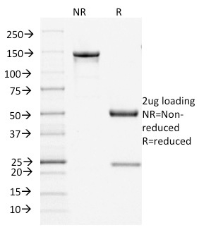
Buffer and Stabilizer: 10mM PBS with 0.05% BSA & 0.05% azide. Also available without BSA & Azide.
Antibody Concentration: 200 ug/ml
Antibody Purification Method:Protein A/G purified from Bioreactor concentrate.
Immunogen: Recombinant human Filaggrin protein fragment (apprx. aa 998-1104) (Please call for additional information.)
Storage Conditions: Store at 2 to 8 C (refrigerate). Stable for 24 months when properly stored.

Filaggrin (Keratinocyte Differentiation Marker) Previously Observed Antibody Staining Patterns

Observed Subcellular, Organelle Specific Staining Data:

Anti-FLG antibody staining is expected to be primarily localized to the vesicles.

Observed Antibody Staining Data By Tissue Type:

Variations in Filaggrin antibody staining intensity in immunohistochemistry on tissue sections are present across different anatomical locations. An intense signal was observed in epidermal cells in the skin. More moderate antibody staining intensity was present in epidermal cells in the skin. Low, but measureable presence of Filaggrin could be seen infibroblasts in skin, melanocytes in skin, squamous epithelial cells in the cervix and uterine. We were unable to detect Filaggrin in other tissues. Disease states, inflammation, and other physiological changes can have a substantial impact on antibody staining patterns. These measurements were all taken in tissues deemed normal or from patients without known disease.

Limitations and Warranty

enQuire Bio's product, Filaggrin (Keratinocyte Differentiation Marker) MonoSpecific Antibody, is available for Research Use Only (RUO-Only). This antibody is guaranteed to work for a period of two years when properly stored.

There are no reviews yet.

Be the first to review “Filaggrin (Keratinocyte Differentiation Marker) Ultraspecific Antibody

Tested against >20,000 Human Proteins

Your email address will not be published. Required fields are marked *

Share a Protocol (View Reviewed Protocols Below)

Protocol Title

Tested against >20,000 Human Proteins

), and application (e.g. IHC, Flow Cytometry, Western Blot) in your title."/>

Materials

Methods

Notes


  protocol submission gif
enQuire Bio, LLC   8420 S Continental Divide Rd, #202   Littleton, CO 80127
Chat: See lower right corner. | enquire@enquirebio.com | Fax: 1-720-897-3730
Customer Service Available M-F 8AM-6PM MST or 24/7 Via Email

Filaggrin (Keratinocyte Differentiation Marker) Ultraspecific Antibody

Tested against >20,000 Human Proteins

https://enquirebio.com/wp-content/uploads/2018/06/enQuire-Bio-2312-MSM5-P0-anti-Filaggrin-Keratinocyte-Differentiation-Marker-antibody-1-300x120.jpeghttps://enquirebio.com/wp-content/uploads/2018/06/enQuire-Bio-2312-MSM5-P0-anti-Filaggrin-Keratinocyte-Differentiation-Marker-antibody-252x300.jpeg