
.jpg)
.jpg)
 PDF Datasheet
Human, Monkey, Mouse (-), and Rat (-) Anti-Bax Antibody Product Attributes
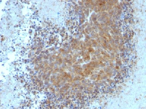
Bax Previously Observed Antibody Staining Patterns
Observed Subcellular, Organelle Specific Staining Data:
Variations in Bax antibody staining intensity in immunohistochemistry on tissue sections are present across different anatomical locations. An intense signal was observed in cells in the seminiferous ducts in testis, cells in the tubules in kidney, epidermal cells in the skin, exocrine glandular cells in the pancreas, glandular cells in the adrenal gland, appendix, breast, cervix, uterine, colon, duodenum, endometrium, gallbladder, parathyroid gland, prostate, rectum, small intestine, stomach and thyroid gland, Leydig cells in the testis, macrophages in lung, respiratory epithelial cells in the bronchus and nasopharynx, trophoblastic cells in the placenta and urothelial cells in the urinary bladder. More moderate antibody staining intensity was present in cells in the seminiferous ducts in testis, cells in the tubules in kidney, epidermal cells in the skin, exocrine glandular cells in the pancreas, glandular cells in the adrenal gland, appendix, breast, cervix, uterine, colon, duodenum, endometrium, gallbladder, parathyroid gland, prostate, rectum, small intestine, stomach and thyroid gland, Leydig cells in the testis, macrophages in lung, respiratory epithelial cells in the bronchus and nasopharynx, trophoblastic cells in the placenta and urothelial cells in the urinary bladder. Low, but measureable presence of Bax could be seen inadipocytes in breast and mesenchymal tissue, cells in the endometrial stroma in endometrium, cells in the granular layer in cerebellum, cells in the molecular layer in cerebellum, follicle cells in the ovary, glial cells in the caudate nucleus, cerebral cortex and hippocampus, hepatocytes in liver, Langerhans in skin, lymphoid tissue in appendix, myocytes in skeletal muscle, neuropil in cerebral cortex, non-germinal center cells in the tonsil, ovarian stroma cells in the ovary, peripheral nerve in mesenchymal tissue, peripheral nerve/ganglion in colon, pneumocytes in lung, smooth muscle cells in the smooth muscle and squamous epithelial cells in the vagina. We were unable to detect Bax in other tissues. Disease states, inflammation, and other physiological changes can have a substantial impact on antibody staining patterns. These measurements were all taken in tissues deemed normal or from patients without known disease.
Observed Antibody Staining Data By Tissue Type:
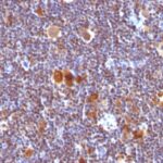
Tissues from cancer patients, for instance, have their own distinct pattern of Bax expression as measured by anti-Bax antibody immunohistochemical staining. The average level of expression by tumor is summarized in the table below. The variability row represents patient to patient variability in IHC staining.
| Sample Type | breast cancer | carcinoid | cervical cancer | colorectal cancer | endometrial cancer | glioma | head and neck cancer | liver cancer | lung cancer | lymphoma | melanoma | ovarian cancer | pancreatic cancer | prostate cancer | renal cancer | skin cancer | stomach cancer | testicular cancer | thyroid cancer | urothelial cancer |
|---|---|---|---|---|---|---|---|---|---|---|---|---|---|---|---|---|---|---|---|---|
| Signal Intensity | ++ | ++ | ++ | ++ | ++ | ++ | ++ | ++ | ++ | ++ | +++ | ++ | ++ | ++ | ++ | ++ | ++ | ++ | ++ | ++ |
| BAX Variability | ++ | ++ | ++ | + | ++ | ++ | ++ | ++ | + | ++ | ++ | ++ | ++ | ++ | ++ | + | ++ | ++ | ++ | + |
| Bax General Information | |
|---|---|
| Alternate Names | |
| Apoptosis regulator BAX, BCL-2-like protein 4, Bcl-2-associated X protein | |
| Molecular Weight | |
| 21kDa | |
| Chromosomal Location | |
| 19q13.33 | |
| Curated Database and Bioinformatic Data | |
| Gene Symbol | BAX |
| Entrez Gene ID | 581 |
| Ensemble Gene ID | ENSG00000087088 |
| RefSeq Protein Accession(s) | XP_016882566, NP_001278358, NP_620119, NP_004315, NP_001278360, NP_620118, NP_001278357, NP_620116, NP_001278359 |
| RefSeq mRNA Accession(s) | XM_017027077 NM_001291430, NM_004324, NM_001291431, NM_138763, NM_001291429, NM_001291428, NM_138764, NR_027882, NM_138761, NM_138762 |
| RefSeq Genomic Accession(s) | NC_018930, NC_000019, NG_012191 |
| UniProt ID(s) | Q5ZPJ1, I6LPK7, Q07812, Q5ZPJ0 |
| UniGene ID(s) | Q5ZPJ1, I6LPK7, Q07812, Q5ZPJ0 |
| HGNC ID(s) | 959 |
| Cosmic ID(s) | BAX |
| KEGG Gene ID(s) | hsa:581 |
| PharmGKB ID(s) | PA25269 |
| General Description of Bax. | |
| Recognizes a protein of 21kDa, identified as the Bax protein. This MAb is highly specific to Bax, shows no cross-reaction with Bcl-2 or Bcl-X protein. Bcl-2 blocks cell death following a variety of stimuli. Bax has extensive amino acid homology with Bcl-2, it homodimerizes, forms heterodimers with Bcl-2. Overexpression of Bax accelerates apoptotic death induced by cytokine deprivation in an IL-3 dependent cell line,, Bax also counters the death repressor activity of Bcl-2. | |
-150x150.jpg)

There are no reviews yet.