Human Anti-CD11c Antibody Product Attributes
CD11c Previously Observed Antibody Staining Patterns
Observed Antibody Staining Data By Tissue Type:
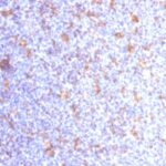
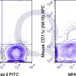
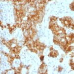
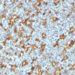
Variations in CD11c antibody staining intensity in immunohistochemistry on tissue sections are present across different anatomical locations. Low, but measureable presence of CD11c could be seen inhematopoietic cells in the bone marrow, lymphoid tissue in appendix, macrophages in lung and squamous epithelial cells in the tonsil. We were unable to detect CD11c in other tissues. Disease states, inflammation, and other physiological changes can have a substantial impact on antibody staining patterns. These measurements were all taken in tissues deemed normal or from patients without known disease.
| CD11c General Information | |
|---|---|
| Alternate Names | |
| Integrin alpha X, ITGAX | |
| Curated Database and Bioinformatic Data | |
| Gene Symbol | ITGAX |
| Entrez Gene ID | 3687 |
| Ensemble Gene ID | ENSG00000140678 |
| RefSeq Protein Accession(s) | XP_011544156, NP_000878, NP_001273304, XP_011544154 |
| RefSeq mRNA Accession(s) | NM_001286375, NM_000887 XR_950797, XM_011545852, XM_011545854 |
| RefSeq Genomic Accession(s) | NG_011451, NC_000016, NC_018927 |
| UniProt ID(s) | H3BN02, P20702 |
| UniGene ID(s) | H3BN02, P20702 |
| HGNC ID(s) | 6152 |
| Cosmic ID(s) | ITGAX |
| KEGG Gene ID(s) | hsa:3687 |
| PharmGKB ID(s) | PA29952 |
| General Description of CD11c. | |
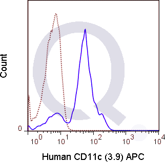
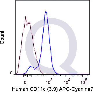
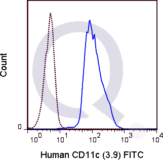
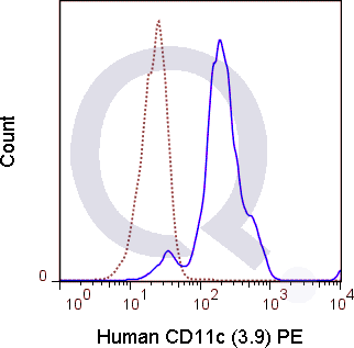
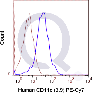
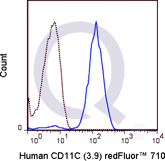
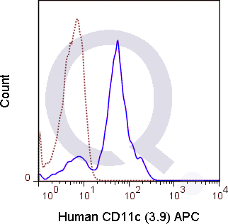
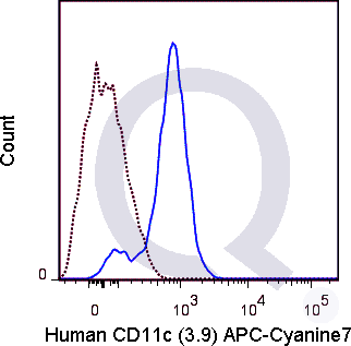
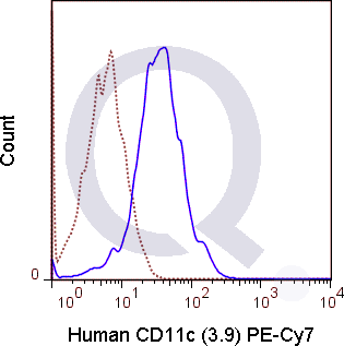
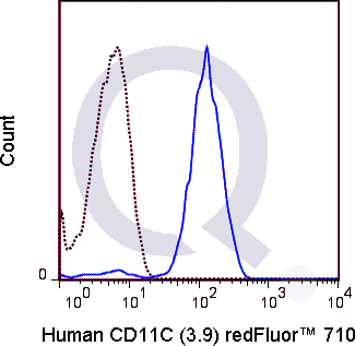
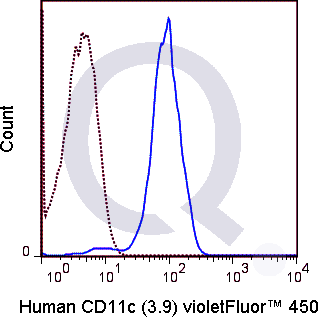
| The 3.9 antibody reacts with human CD11c, also known as integrin alpha X. This 150 kDa cell surface glycoprotein is part of a family of integrin receptors that mediate adhesion between cells (cell-cell) and components of the extracellular matrix, e.g. fibrinogen (cell-matrix). In addition, integrins are active signaling receptors which recruit leukocytes to inflammatory sites and promote cell activation. Complete, functional integrin receptors consist of distinct combinations of integrin chains which are differentially expressed. Integrin alpha X (CD11c) assembles with Integrin beta-2 (CD18) into a receptor known as CR4 which can bind and induce signaling through ICAMs and VCAM-1 on endothelial cells and can also facilitate removal of iC3b bearing foreign cells.The 3.9 antibody is widely used as a marker for CD11c expression on dendritic cells (DC), often in parallel with markers for CD11b, for identification of developmental stages and mature subsets of this cell type. CD11c is prominently expressed on tissue macrophages, and is also detected on activated neutrophils, granulocytes, some types of activated T cells and intestinal intraepithelial lymphocytes (IEL). The antibody is reported to be cross-reactive with Baboon, Chimpanzee, Cynomolgus and Rhesus CD11c. | |
Selected References
Limitations and Warranty
| Size | |
|---|---|
| Tag | APC, APC-Cy7, FITC, PE, PE-Cy7, Qfluor™ 630, Qfluor™ 710, Unconjugated, V450 |
| Buffer and Stabilizer | 10 mM NaH2PO4, 150 mM NaCl, 0.09% NaN3, 0.1% gelatin, pH7.2, 10 mM NaH2PO4, 150 mM NaCl, 0.09% NaN3, pH 7.2 |
| Product Type | |
| Host | |
| Isotype | |
| Applications | |
| Species | |
| Mass Spec Validated? |
Only logged in customers who have purchased this product may leave a review.





There are no reviews yet.