

 PDF Datasheet
Human Anti-CD14 Antibody Product Attributes
CD14 Previously Observed Antibody Staining Patterns
Observed Subcellular, Organelle Specific Staining Data:
Staining with anti-CD14 antibody reveals CD14 expression is expected to be primarily localized to the plasma membrane and vesicles.
Observed Antibody Staining Data By Tissue Type:
Variations in CD14 antibody staining intensity in immunohistochemistry on tissue sections are present across different anatomical locations. An intense signal was observed in endothelial cells in the colon. More moderate antibody staining intensity was present in endothelial cells in the colon. Low, but measureable presence of CD14 could be seen in cells in the seminiferous ducts in testis, fibroblasts in skin, glandular cells in the fallopian tube and salivary gland, Langerhans in skin and Leydig cells in the testis. We were unable to detect CD14 in other tissues. Disease states, inflammation, and other physiological changes can have a substantial impact on antibody staining patterns. These measurements were all taken in tissues deemed normal or from patients without known disease.
Observed Antibody Staining Data By Tissue Disease Status:
Tissues from cancer patients, for instance, have their own distinct pattern of CD14 expression as measured by anti-CD14 antibody immunohistochemical staining. The average level of expression by tumor is summarized in the table below. The variability row represents patient to patient variability in IHC staining.
| Sample Type | breast cancer | carcinoid | cervical cancer | colorectal cancer | endometrial cancer | glioma | head and neck cancer | liver cancer | lung cancer | lymphoma | melanoma | ovarian cancer | pancreatic cancer | prostate cancer | renal cancer | skin cancer | stomach cancer | testicular cancer | thyroid cancer | urothelial cancer |
|---|---|---|---|---|---|---|---|---|---|---|---|---|---|---|---|---|---|---|---|---|
| Signal Intensity | – | – | – | – | – | – | – | – | – | – | – | – | – | – | – | – | – | – | – | – |
| CD14 Variability | + | ++ | + | + | + | + | + | ++ | + | + | + | ++ | ++ | + | + | + | + | + | + | + |
| CD14 General Information | |
|---|---|
| Alternate Names | |
| mCD14 | |
| Curated Database and Bioinformatic Data | |
| Gene Symbol | CD14 |
| Entrez Gene ID | 929 |
| Ensemble Gene ID | ENSG00000170458 |
| RefSeq Protein Accession(s) | NP_001035110, NP_001167575, NP_001167576, NP_000582 |
| RefSeq mRNA Accession(s) | NM_001174104, NM_001040021, NM_000591, NM_001174105 |
| RefSeq Genomic Accession(s) | NG_023178, NC_018916, NC_000005 |
| UniProt ID(s) | P08571 |
| UniGene ID(s) | P08571 |
| HGNC ID(s) | 1628 |
| Cosmic ID(s) | CD14 |
| KEGG Gene ID(s) | hsa:929 |
| PharmGKB ID(s) | PA26188 |
| General Description of CD14. | |
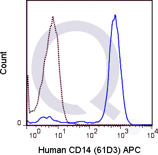
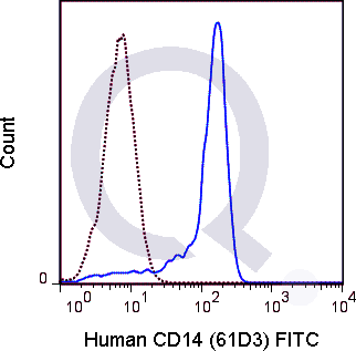
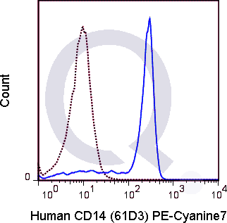
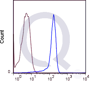
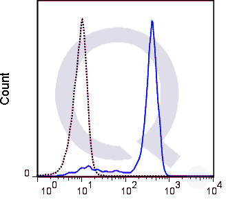
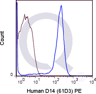
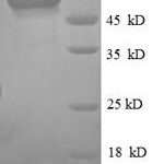
| Anti-human CD14 antibody, clone 61D3, is a widely used, highly published anti-CD14 clone. It binds a ~40 kDa glycoprotein identified as CD14. In the literature, the principle use of this antibody is in flow cytometry. CD14 is highly expressed on monocytes and is also present on neutrophils, several populations of macrophages, and some DCs. It may be membrane bound but is also secreted as sCD14. CD14 cooperates with TLR4 and MD2 in mediating the innate immune response to the lipopolysaccharide (LPS) which drives NFkB activation. Although best known for binding LPS, CD14 can also bind lipoteichoic acid. | |

Kang et al.
Flow Cytometry with anti-CD14 (61D3) of human cells from gastric cancer samples.
Materials
Methods
Notes
This protocol details procedures used in the Kang lab at the Sungkyunkwan University School of Medicine and published in Choi, H. S. et al. The prognostic effects of tumor infiltrating regulatory T cells and myeloid derived suppressor cells assessed by multicolor flow cytometry in gastric cancer patients. Oncotarget 7, 7940–51 (2016).