Human Anti-CD3 Antibody Product Attributes
CD3 Previously Observed Antibody Staining Patterns
Observed Antibody Staining Data By Tissue Type:
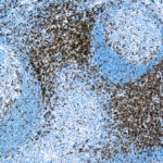
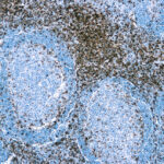
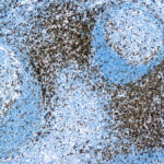
Variations in CD3 antibody staining intensity in immunohistochemistry on tissue sections are present across different anatomical locations. An intense signal was observed in lymphoid tissue in appendix and non-germinal center cells in the lymph node and tonsil. More moderate antibody staining intensity was present in lymphoid tissue in appendix and non-germinal center cells in the lymph node and tonsil. Low, but measureable presence of CD3 could be seen in. We were unable to detect CD3 in other tissues. Disease states, inflammation, and other physiological changes can have a substantial impact on antibody staining patterns. These measurements were all taken in tissues deemed normal or from patients without known disease.
| CD3 General Information | |
|---|---|
| Alternate Names | |
| Leu-4, T3 | |
| Curated Database and Bioinformatic Data | |
| Gene Symbol | CD3E |
| Entrez Gene ID | 916 |
| Ensemble Gene ID | ENSG00000198851 |
| RefSeq Protein Accession(s) | NP_000724 |
| RefSeq mRNA Accession(s) | NM_000733, |
| RefSeq Genomic Accession(s) | NC_018922, NG_007383, NC_000011 |
| UniProt ID(s) | P07766 |
| UniGene ID(s) | P07766 |
| HGNC ID(s) | 1674 |
| Cosmic ID(s) | CD3E |
| KEGG Gene ID(s) | hsa:916 |
| PharmGKB ID(s) | PA26216 |
| General Description of CD3. | |
| The OKT3 antibody is specific for human CD3e, also known as CD3 epsilon, a 20 kDa subunit of the T cell receptor complex along with CD3 gamma and CD3 delta. These integral membrane protein chains assemble with additional chains of the T cell receptor (TCR), as well as CD3 zeta chain, to form the T cell receptor – CD3 complex. Together with co-receptors CD4 or CD8, the complex serves to recognize antigens bound to MHC molecules on antigen-presenting cells. These interactions promote T cell receptor signaling (T cell activation), inducing a number of cellular responses including proliferation, differentiation, production of cytokines or activation-induced cell death. CD3 is differentially expressed during thymocyte-to-T cell development and on all mature T cells.The OKT3 antibody is a widely used phenotypic marker for human T cells. In addition, as the CD3e subunit of the TCR complex contains intracellular signaling domains, binding of OKT3 antibody to CD3e can induce cell activation. A recent publication of the crystal structure of a CD3e-mitogenic antibody complex provides further insight as to the action of commonly used agonist antibodies, as well as specific epitope-binding data for the widely used human CD3 antibodies OKT3 and UCHT1 (Fernandes, R.A. et al. 2012. J. Biol. Chem. 287: 13324-13335). OKT3 has also been shown to be cross-reactive with Chimpanzee CD3 and has been used for in vitro activation of T cells in this species. Please choose the appropriate format for each application. | |
Selected References
Limitations and Warranty
| Size | |
|---|---|
| Tag | APC, FITC, PE, PE-Cy7, PerCP-Cy5.5, Qfluor™ 630, Qfluor™ 710, Unconjugated, V450 |
| Buffer and Stabilizer | 10 mM NaH2PO4, 150 mM NaCl, 0.09% NaN3, 0.1% gelatin, pH7.2, 10 mM NaH2PO4, 150 mM NaCl, 0.09% NaN3, pH 7.2 |
| Product Type | |
| Host | |
| Isotype | |
| Applications | |
| Species | |
| Mass Spec Validated? |
Only logged in customers who have purchased this product may leave a review.

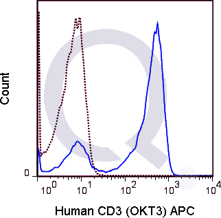

![Anti-CD3 Antibody [OKT3] - Image 3](https://cdn-enquirebio.pressidium.com/wp-content/uploads/2017/10/enQuire-Bio-QAB5-F-100Tests-anti-CD3-antibody-10.png)
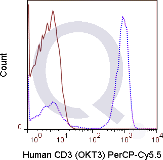
![Human PBMCs were stained with 5 uL PE conjugated anti-CD3 antibody [OKT3] (solid line) or 0.5 ug PE Mouse IgG2a isotype control (dashed line). Flow Cytometry Data from 10,000 events.](https://cdn-enquirebio.pressidium.com/wp-content/uploads/2017/10/enQuire-Bio-QAB5-PE-100Tests-anti-CD3-antibody-10.png)
![Human PBMCs were stained with 5 uL of PE-Cy7 anti-human CD3 antibody [OKT-3] antibody and analyzed via flow cytometry.](https://cdn-enquirebio.pressidium.com/wp-content/uploads/2017/10/enQuire-Bio-QAB5-PE7-100Tests-anti-CD3-antibody-10.png)
![Human PBMCs were stained with 5 uL Qfluor™ 710 conjugated anti-human CD3 antibody [clone OKT3] (solid line) or 1 ug Qfluor™ 710 Mouse IgG2a isotype control (dashed line). Flow Cytometry Data from 10,000 events.](https://cdn-enquirebio.pressidium.com/wp-content/uploads/2017/10/enQuire-Bio-QAB5-QF710-100Tests-anti-CD3-antibody-10.png)
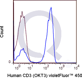
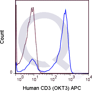
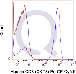
![Human PBMCs were stained with 5 uL PE conjugated anti-CD3 antibody [OKT3] (solid line) or 0.5 ug PE Mouse IgG2a isotype control (dashed line). Flow Cytometry Data from 10,000 events.](https://cdn-enquirebio.pressidium.com/wp-content/uploads/2017/11/enQuire-Bio-QAB5-PE-100Tests-anti-CD3-antibody-9.png)
![Human PBMCs were stained with 5 uL of PE-Cy7 anti-human CD3 antibody [OKT-3] antibody and analyzed via flow cytometry.](https://cdn-enquirebio.pressidium.com/wp-content/uploads/2017/11/enQuire-Bio-QAB5-PE7-100Tests-anti-CD3-antibody-9.png)
![Human PBMCs were stained with 5 uL Qfluor™ 710 conjugated anti-human CD3 antibody [clone OKT3] (solid line) or 1 ug Qfluor™ 710 Mouse IgG2a isotype control (dashed line). Flow Cytometry Data from 10,000 events.](https://cdn-enquirebio.pressidium.com/wp-content/uploads/2017/11/enQuire-Bio-QAB5-QF710-100Tests-anti-CD3-antibody-9.png)

There are no reviews yet.