

 PDF Datasheet
Human Anti-CD54 / ICAM-1 Antibody Product Attributes
CD54 / ICAM-1 Previously Observed Antibody Staining Patterns
Observed Antibody Staining Data By Tissue Type:
Variations in CD54 / ICAM-1 antibody staining intensity in immunohistochemistry on tissue sections are present across different anatomical locations. An intense signal was observed in cells in the glomeruli in kidney, endothelial cells in the colon and pneumocytes in lung. More moderate antibody staining intensity was present in cells in the glomeruli in kidney, endothelial cells in the colon and pneumocytes in lung. Low, but measureable presence of CD54 / ICAM-1 could be seen in cells in the seminiferous ducts in testis, germinal center cells in the lymph node, glandular cells in the fallopian tube and urothelial cells in the urinary bladder. We were unable to detect CD54 / ICAM-1 in other tissues. Disease states, inflammation, and other physiological changes can have a substantial impact on antibody staining patterns. These measurements were all taken in tissues deemed normal or from patients without known disease.
Observed Antibody Staining Data By Tissue Disease Status:
Tissues from cancer patients, for instance, have their own distinct pattern of CD54 / ICAM-1 expression as measured by anti-CD54 / ICAM-1 antibody immunohistochemical staining. The average level of expression by tumor is summarized in the table below. The variability row represents patient to patient variability in IHC staining.
| Sample Type | breast cancer | carcinoid | cervical cancer | colorectal cancer | endometrial cancer | glioma | head and neck cancer | liver cancer | lung cancer | lymphoma | melanoma | ovarian cancer | pancreatic cancer | prostate cancer | renal cancer | skin cancer | stomach cancer | testicular cancer | thyroid cancer | urothelial cancer |
|---|---|---|---|---|---|---|---|---|---|---|---|---|---|---|---|---|---|---|---|---|
| Signal Intensity | – | – | – | – | – | – | + | – | – | – | – | + | – | – | – | – | – | – | – | – |
| ICAM1 Variability | ++ | + | ++ | + | + | + | ++ | ++ | ++ | + | ++ | ++ | + | + | ++ | ++ | + | + | + | + |
| CD54 / ICAM-1 General Information | |
|---|---|
| Alternate Names | |
| ICAM-1, Intercellular Adhesion Molecule 1, CD54, Cluster of Differentiation 54, ICAM1 | |
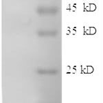
| Molecular Weight | |
| 85-115kDa | |
| Chromosomal Location | |
| 19p13.3-p13.2 | |
| Curated Database and Bioinformatic Data | |
| Gene Symbol | ICAM1 |
| Entrez Gene ID | 3383 |
| Ensemble Gene ID | ENSG00000090339 |
| RefSeq Protein Accession(s) | NP_000192 |
| RefSeq mRNA Accession(s) | NM_000201 |
| RefSeq Genomic Accession(s) | NC_000019, NG_012083, NC_018930 |
| UniProt ID(s) | P05362 |
| UniGene ID(s) | P05362 |
| HGNC ID(s) | 5344 |
| Cosmic ID(s) | ICAM1 |
| KEGG Gene ID(s) | hsa:3383 |
| PharmGKB ID(s) | PA29592 |
| General Description of CD54 / ICAM-1. | |
| Recognizes an 85-115kDa protein (variation with cell type), identified as intercellular adhesion molecule (ICAM-1) (Workshop IV). It has 7 potential N-linked glycosylation sites. ICAM-1 is a single chain glycoprotein of Ig supergene family, present on unstimulated endothelial cells (EC), on a variety of other cell types including activated fibroblasts, EC, macrophages,, lymphocytes. ICAM-1 mediates cell adhesion by binding to integrins CD11a/CD18 (leukocyte adhesion molecule, LFA-1), to CD11b/CD18 (Mac-1). This interaction enhances antigen-specific T-cell activation. ICAM-1 also binds to CD43, to Plasmodium falciparum infected RBCs. W-CAM-1 MAb blocks aggregation of cell lines mediated by the ICAM-1, blocks homotypic binding of purified populations of activated T-, B-lymphocytes, also aggregation of mixed T-, B-cell blasts. It inhibits T-cell adhesion to normal human endothelial cells. Activation induced by cell-cell contact (mixed lymphocyte reaction, T-cell mediated B-cell activation) is significantly inhibited. This MAb blocks elements of both effector arms of immune system (cytotoxic cell function, Ig production). | |
There are no reviews yet.