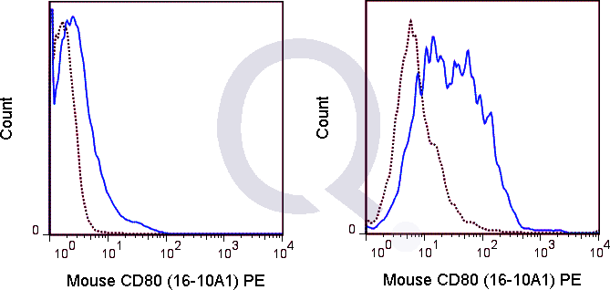
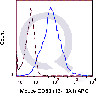
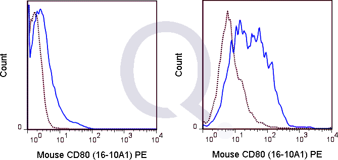
Mouse Anti-CD80 Antibody Product Attributes
CD80 Previously Observed Antibody Staining Patterns
Observed Antibody Staining Data By Tissue Type:
Variations in CD80 antibody staining intensity in immunohistochemistry on tissue sections are present across different anatomical locations. Low, but measureable presence of CD80 could be seen in. We were unable to detect CD80 in other tissues. Disease states, inflammation, and other physiological changes can have a substantial impact on antibody staining patterns. These measurements were all taken in tissues deemed normal or from patients without known disease.
| CD80 General Information | |
|---|---|
| Alternate Names | |
| B7-1, B7.1 | |
| Curated Database and Bioinformatic Data | |
| Gene Symbol | Cd80 |
| Entrez Gene ID | 12519 |
| Ensemble Gene ID | ENSMUSG00000075122 |
| RefSeq Protein Accession(s) | XP_017172346, NP_033985, XP_006521800, XP_017172347 |
| RefSeq mRNA Accession(s) | NM_009855, XM_017316857, XM_017316858, XM_006521737 |
| RefSeq Genomic Accession(s) | NC_000082 |
| UniProt ID(s) | Q00609, Q549R2 |
| UniGene ID(s) | Q00609, Q549R2 |
| Cosmic ID(s) | Cd80 |
| KEGG Gene ID(s) | mmu:12519 |
| General Description of CD80. | |
| The 16-10A1 antibody reacts with mouse CD80, also known as B7-1, a 55 kDa type I transmembrane protein ligand for CD152 (CTLA-4) and for CD28, a co-stimulatory receptor for the T cell receptor (TCR). CD28 also binds a second B7 ligand known as CD86 (B7-2). Both CD80 and CD86 are expressed on activated B cells and antigen-presenting cells. These ligands trigger CD28 signaling in concert with TCR activation to drive T cell proliferation, induce high-level expression of IL-2, impart resistance to apoptosis, and enhance T cell cytotoxicity. The interaction / co-stimulatory signaling between the B7 ligands and CD28 or CTLA-4 provides crucial communication between T cells and B cells or APCs to coordinate the adaptive immune response. | |


![Anti-CD80 Antibody [16-10A1] - Image 3](https://cdn-enquirebio.pressidium.com/wp-content/uploads/2017/10/enQuire-Bio-QAB50-F-100ug-anti-CD80-antibody-10.png)


There are no reviews yet.