
.jpg)
 PDF Datasheet
Human Anti-Wilm’s Tumor 1 Antibody Product Attributes
Wilm’s Tumor 1 Previously Observed Antibody Staining Patterns
Observed Subcellular, Organelle Specific Staining Data:
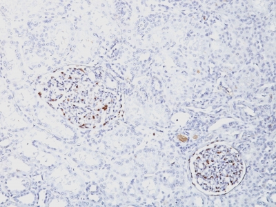
Anti-WT1 antibody staining is expected to be primarily localized to the nucleoplasm.
Observed Antibody Staining Data By Tissue Type:
Variations in Wilm’s Tumor 1 antibody staining intensity in immunohistochemistry on tissue sections are present across different anatomical locations. An intense signal was observed in myoepithelial cells in the breast, cells in the endometrial stroma in endometrium, glandular cells in the fallopian tube, cells in the glomeruli in kidney and cells in the seminiferous ducts in testis. More moderate antibody staining intensity was present in myoepithelial cells in the breast, cells in the endometrial stroma in endometrium, glandular cells in the fallopian tube, cells in the glomeruli in kidney and cells in the seminiferous ducts in testis. Low, but measureable presence of Wilm’s Tumor 1 could be seen inglandular cells in the breast and epididymis, cells in the tubules in kidney, decidual cells in the placenta, trophoblastic cells in the placenta, glandular cells in the prostate and seminal vesicle, adipocytes in mesenchymal tissue, fibroblasts in mesenchymal tissue and cells in the red pulp in spleen. We were unable to detect Wilm’s Tumor 1 in other tissues. Disease states, inflammation, and other physiological changes can have a substantial impact on antibody staining patterns. These measurements were all taken in tissues deemed normal or from patients without known disease.
Observed Antibody Staining Data By Tissue Disease Status:
Tissues from cancer patients, for instance, have their own distinct pattern of Wilm’s Tumor 1 expression as measured by anti-Wilm’s Tumor 1 antibody immunohistochemical staining. The average level of expression by tumor is summarized in the table below. The variability row represents patient to patient variability in IHC staining.
| Sample Type | breast cancer | carcinoid | cervical cancer | colorectal cancer | endometrial cancer | glioma | head and neck cancer | liver cancer | lung cancer | lymphoma | melanoma | ovarian cancer | pancreatic cancer | prostate cancer | renal cancer | skin cancer | stomach cancer | testicular cancer | thyroid cancer | urothelial cancer |
|---|---|---|---|---|---|---|---|---|---|---|---|---|---|---|---|---|---|---|---|---|
| Signal Intensity | – | – | – | – | – | +++ | – | – | – | – | ++ | +++ | – | – | – | – | – | – | – | – |
| WT1 Variability | ++ | + | + | + | + | ++ | + | + | + | + | ++ | ++ | + | + | ++ | + | + | + | + | ++ |
| Wilm’s Tumor 1 General Information | |
|---|---|
| Alternate Names | |
| Wilms tumor protein WT1, Wilms tumor antigen, Wilms Tumor 1 | |
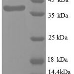
| Molecular Weight | |
| 47-55kDa | |
| Chromosomal Location | |
| 11p13 | |
| Curated Database and Bioinformatic Data | |
| Gene Symbol | WT1 |
| Entrez Gene ID | 7490 |
| Ensemble Gene ID | ENSG00000184937 |
| RefSeq Protein Accession(s) | NP_077742, NP_001185481, NP_077744, NP_000369, NP_001185480 |
| RefSeq mRNA Accession(s) | NM_024426, NM_024425, NM_001198551, NM_001198552, NM_024424 NM_000378 |
| RefSeq Genomic Accession(s) | NC_018922, NC_000011, NG_009272, |
| UniProt ID(s) | A0A1W2PQQ0, Q6PI38, P19544, A0A1W2PR07, A0A1W2PPP2 |
| UniGene ID(s) | A0A1W2PQQ0, Q6PI38, P19544, A0A1W2PR07, A0A1W2PPP2 |
| HGNC ID(s) | 12796 |
| Cosmic ID(s) | WT1 |
| KEGG Gene ID(s) | hsa:7490 |
| PharmGKB ID(s) | PA37395 |
| General Description of Wilm’s Tumor 1. | |
| Recognizes a 47-55kDa-tumor suppressor protein, identified as Wilm’s Tumor (WT1) protein. The antibody reacts with all isoforms of the full-length WT1 and also identifies WT1 lacking exon 2-encoded amino acids, frequently found in subsets of sporadic Wilm s tumors. WT1, a sporadic and familial pediatric kidney tumor, is genetically heterogeneous. Wilm s tumor is associated with mutations of WT1, a zinc-finger transcription factor that is essential for the development of the metanephric kidney and the urogenital system. The WT1 gene is normally expressed in fetal kidney and mesothelium, and its expression has been suggested as a marker for Wilm s tumor and mesothelioma. WT1 protein has been identified in proliferative mesothelial cells, malignant mesothelioma, ovarian carcinoma, gonadoblastoma, nephroblastoma, and desmoplastic small round cell tumor. Lung adenocarcinomas rarely stain positive with this antibody. WT1 protein expression in mesothelial cells has become a reliable marker for the diagnosis of mesotheliomas. | |
-150x150.jpg)

-150x150.jpg)
-150x150.jpg)
-150x150.jpg)
There are no reviews yet.