

Human Anti-CD54 / ICAM-1 Antibody Product Attributes
Species: Human
Cross Reactivity: pig
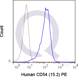
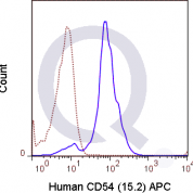
Tested Applications: Flow Cytometry.
Application Notes: 5 ul per test where one test represents staining of a cell sample in a final volume of approximately 100 uL. The number of cells within a sample must be experimentally determined, but ranges between 1x10⁵ to 1x10⁸ cells.
Clonality: Monoclonal Antibody
Anti-CD54 / ICAM-1 Antibody Clone: 15.2
Clone 15.2 Host and Isotype: Mouse IgG1
Buffer and Stabilizer: 10 mM NaH2PO4, 150 mM NaCl, 0.09% NaN3, 0.1% gelatin, pH7.2
Antibody Concentration: 5 uL /test
Storage Conditions: 2-8C protected from light. Stable for 12 Months. Do Not Freeze.
CD54 / ICAM-1 Previously Observed Antibody Staining Patterns
Observed Antibody Staining Data By Tissue Type:
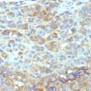
Variations in CD54 / ICAM-1 antibody staining intensity in immunohistochemistry on tissue sections are present across different anatomical locations. An intense signal was observed in cells in the glomeruli in kidney, endothelial cells in the colon and pneumocytes in lung. More moderate antibody staining intensity was present in cells in the glomeruli in kidney, endothelial cells in the colon and pneumocytes in lung. Low, but measureable presence of CD54 / ICAM-1 could be seen in cells in the seminiferous ducts in testis, germinal center cells in the lymph node, glandular cells in the fallopian tube and urothelial cells in the urinary bladder. We were unable to detect CD54 / ICAM-1 in other tissues. Disease states, inflammation, and other physiological changes can have a substantial impact on antibody staining patterns. These measurements were all taken in tissues deemed normal or from patients without known disease.Observed Antibody Staining Data By Tissue Disease Status:
Tissues from cancer patients, for instance, have their own distinct pattern of CD54 / ICAM-1 expression as measured by anti-CD54 / ICAM-1 antibody immunohistochemical staining. The average level of expression by tumor is summarized in the table below. The variability row represents patient to patient variability in IHC staining.| Sample Type | breast cancer | carcinoid | cervical cancer | colorectal cancer | endometrial cancer | glioma | head and neck cancer | liver cancer | lung cancer | lymphoma | melanoma | ovarian cancer | pancreatic cancer | prostate cancer | renal cancer | skin cancer | stomach cancer | testicular cancer | thyroid cancer | urothelial cancer |
|---|---|---|---|---|---|---|---|---|---|---|---|---|---|---|---|---|---|---|---|---|
| Signal Intensity | - | - | - | - | - | - | + | - | - | - | - | + | - | - | - | - | - | - | - | - |
| ICAM1 Variability | ++ | + | ++ | + | + | + | ++ | ++ | ++ | + | ++ | ++ | + | + | ++ | ++ | + | + | + | + |
Selected References
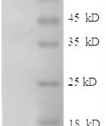
Sommaggio R, Cohnen A, Watzl C, and Costa C. 2012. J. Immunol. 188: 2075-2083. (in vitro blocking - Pig)Avril M, Tripathi AK, Brazier AJ, Andisi C, Janes JH, Soma VL, Sullivan DJ, Bull PC, Stins MF, and Smith JD. 2012. Proc. Natl. Acad. Sci. 109: E1782-E1790. (in vitro blocking)Dryden NH, Sperone A, Martin-Almedina S, Hannah RL, Birdsey GM, Khan ST, Layhadi JA et al. 2012. J. Biol. Chem. 287: 12331-12342. (Western Blot)Di Lorenzo A, Manes TD, Davalos A, Wright PL, and Sessa WC. 2011. Blood. 117: 2284-2295. (in vitro activation/cross-linking)Kim S, and Nadel JA. 2009. Am. J. Physiol. Lung Cell. Mol. Physiol. 297: L174-L183. (in vitro blocking, Western Blot)Goto E, Kohrogi H, Hirata N, Tsumori K, Hirosako S, Hamamoto J, Fujii K, Kawano O, and Ando M. 2000. Am. J. Respir. Cell Mol. Biol. 22: 405-411. (Immunohistochemistry - frozen tissue)
Limitations and Warranty
enQuire Bio's Human Anti-CD54 / ICAM-1 Monoclonal is available for Research Use Only. This antibody is guaranteed to work for a period of two years when properly stored.

There are no reviews yet.