Human HLA-DRB Recombinant Protein Product Attributes
Product Type: Recombinant Protein
Recombinant HLA-DRB based upon sequence from: Human
Host: QP8911 protein expressed in E. coli.
Tag: His
Protein Construction: A DNA sequence encoding the Homo sapiens (Human) HLA-DRB, was expressed in the hosts and tags indicated. Please select your host/tag option, above.
Application Notes: Please contact us for application specific information for QP8911.
Bioactivity Data: Untested
Full Length? Full Length
Expression Region: Asp31 – Ser266
Amino Acid Sequence: DTRPRFLEYS TGECYFFNGT ERVRLLERHF HNQEELLRFD SDVGEFRAVT ELGRPVAESW NSQKDILEDR RAAVDTYCRH NYGAVESFTV QRRVHPKVTV YPSKTQPLQH HNLLVCSVSG FYPGSIEVRW FRNGQEEKTG VVSTGLIHNG DWTFQTLVML ETVPRSGEVY TCQVEHPSVT SPLTVEWRAR SESAQSKMLS GVGGFVLGLL FLGAGLFIYF RNQKGHSGLQ PRGFLS
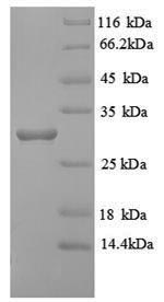
Purity: Greater than 90% as determined by SDS-PAGE.
Reconstitution Instructions:
Concentration of Human HLA-DRB Protein:
Endotoxin Levels: Not determined.
Buffer: Tris-based buffer, 50% glycerol
Storage Conditions: Store at -20C to -80C.
| Recombinant Human HLA-DRB Protein (31-266aa) General Information | |
|---|---|
| Curated Database and Bioinformatic Data | |
| Gene Symbol | HLA-DRB1 |
| Ensemble Gene ID | ENSG00000206306 |
| UniProt ID(s) | Q95IE3 |
| UniGene ID(s) | Hs.534322 |
| HGNC ID(s) | HGNC:4948 |
| COSMIC ID Link(s) | HLA-DRB1 |
| General Description of Recombinant Human HLA-DRB Protein (31-266aa). | |
| Binds peptides derived from antigens that access the endocytic route of antigen presenting cells (APC) and presents th on the cell surface for recognition by the CD4 T-cells. The peptide binding cleft accommodates peptides of 10-30 residues. The peptides presented by MHC class II molecules are generated mostly by degradation of proteins that access the endocytic route, where they are processed by lysosomal proteases and other hydrolases. Exogenous antigens that have been endocytosed by the APC are thus readily available for presentation via MHC II molecules, and for this reason this antigen presentation pathway is usually referred to as exogenous. As mbrane proteins on their way to degradation in lysosomes as part of their normal turn-over are also contained in the endosomal/lysosomal compartments, exogenous antigens must compete with those derived from endogenous components. Autophagy is also a source of endogenous peptides, autophagosomes constitutively fuse with MHC class II loading compartments. In addition to APCs, other cells of the gastrointestinal tract, such as epithelial cells, express MHC class II molecules and CD74 and act as APCs, which is an unusual trait of the GI tract. To produce a MHC class II molecule that presents an antigen, three MHC class II molecules (heterodimers of an alpha and a beta chain) associate with a CD74 trimer in the ER to form a heterononamer. Soon after the entry of this complex into the endosomal/lysosomal syst where antigen processing occurs, CD74 undergoes a sequential degradation by various proteases, including CTSS and CTSL, leaving a small fragment termed CLIP (class-II-associated invariant chain peptide). The roval of CLIP is facilitated by HLA-DM via direct binding to the alpha-beta-CLIP complex so that CLIP is released. HLA-DM stabilizes MHC class II molecules until primary high affinity antigenic peptides are bound. The MHC II molecule bound to a peptide is then transported to the cell mbrane surface. In B-cells, the interaction between HLA-DM and MHC class II molecules is regulated by HLA-DO. Primary dendritic cells (DCs) also to express HLA-DO. Lysosomal microenvironment has been implicated in the regulation of antigen loading into MHC II molecules, increased acidification produces increased proteolysis and efficient peptide loading. | |
Limitations and Performance Guarantee
This is a life science research product (for Research Use Only). This product is guaranteed to work for a period of two years when stored at -70C or colder, and one year when aliquoted and stored at -20C.



There are no reviews yet.