![Analysis of Mass Spec data (dashed-line) of fractions stained with Cytokeratin 7 MS-QAVA™ monoclonal antibody [Clone: KRT7/903] (solid-line), reveals that less than 7.5% of signal is attributable to non-specific binding of anti-Cytokeratin 7 [Clone KRT7/903] to targets other than KRT7 protein. Even frequently cited antibodies have much greater non-specific interactions, averaging over 30%. Data in image is from analysis in A431, RT4 and MCF7 cells.](https://cdn-enquirebio.pressidium.com/wp-content/uploads/2017/10/enQuire-Bio-3855-MSM3-P1-anti-Cytokeratin-7-antibody.png "Cytokeratin 7 Antibody Mass Spec Validation Data")
Human, Mouse (-), Rat (-), Ferret (-), and Rabbit (-) Anti-Cytokeratin 7 Antibody Product Attributes
Species: Human, Mouse (-), Rat (-), Ferret (-), and Rabbit (-)
Tested Applications: Flow Cytometry, Immunofluorescence, Immunohistochemistry (IHC). Additionally, this antibody has been validated by MS-QAVAâ¢. Specificity has been quantitatively determined via Mass Spec.
Clonality: Monoclonal
Anti-Cytokeratin 7 Antibody Clone: KRT7/903
Clone KRT7/903 Host and Isotype: Mouse IgG1
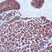
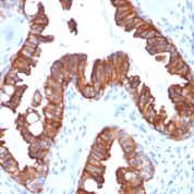
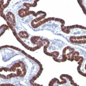
Anti-Human, Mouse (-), Rat (-), Ferret (-), and Rabbit (-) Cytokeratin 7 Positive Control Sample: A431, HeLa cells, Breast cancer
Cellular Localization of Antibody Cytoplasmic
Buffer and Stabilizer: 10mM PBS with 0.05% BSA & 0.05% azide.
Antibody Concentration: 200ug/ml
Antibody Purification Method:Protein A/G Purified
Immunogen: Recombinant full-length human KRT7 protein
Storage Conditions: Store at 2 to 8° C (refrigerate). Stable for 24 months when properly stored.
Cytokeratin 7 Previously Observed Antibody Staining Patterns
Observed Subcellular, Organelle Specific Staining Data:
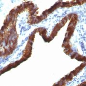
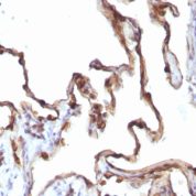
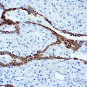
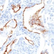
Variations in Cytokeratin 7 antibody staining intensity in immunohistochemistry on tissue sections are present across different anatomical locations. An intense signal was observed in glandular cells in the breast, cervix, uterine, endometrium, fallopian tube, gallbladder, salivary gland, seminal vesicle and thyroid gland, respiratory epithelial cells in the bronchus and nasopharynx, trophoblastic cells in the placenta and urothelial cells in the urinary bladder. More moderate antibody staining intensity was present in glandular cells in the breast, cervix, uterine, endometrium, fallopian tube, gallbladder, salivary gland, seminal vesicle and thyroid gland, respiratory epithelial cells in the bronchus and nasopharynx, trophoblastic cells in the placenta and urothelial cells in the urinary bladder. Low, but measureable presence of Cytokeratin 7 could be seen inglandular cells in the parathyroid gland and stomach and myoepithelial cells in the breast. We were unable to detect Cytokeratin 7 in other tissues. Disease states, inflammation, and other physiological changes can have a substantial impact on antibody staining patterns. These measurements were all taken in tissues deemed normal or from patients without known disease.Observed Antibody Staining Data By Tissue Type:
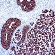
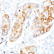
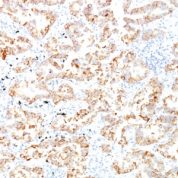
Tissues from cancer patients, for instance, have their own distinct pattern of Cytokeratin 7 expression as measured by anti-Cytokeratin 7 antibody immunohistochemical staining. The average level of expression by tumor is summarized in the table below. The variability row represents patient to patient variability in IHC staining.| Sample Type | breast cancer | carcinoid | cervical cancer | colorectal cancer | endometrial cancer | glioma | head and neck cancer | liver cancer | lung cancer | lymphoma | melanoma | ovarian cancer | pancreatic cancer | prostate cancer | renal cancer | skin cancer | stomach cancer | testicular cancer | thyroid cancer | urothelial cancer |
|---|---|---|---|---|---|---|---|---|---|---|---|---|---|---|---|---|---|---|---|---|
| Signal Intensity | +++ | - | +++ | - | ++ | - | - | + | +++ | - | - | +++ | +++ | + | - | - | +++ | - | +++ | +++ |
| KRT7 Variability | ++ | + | ++ | + | ++ | + | + | ++ | + | + | + | + | + | +++ | + | ++ | ++ | + | + | + |
Limitations and Warranty
This product is for Research Use Only. This antibody is guaranteed to work for a period of two years when properly stored.

![Analysis of Mass Spec data (dashed-line) of fractions stained with Cytokeratin 7 MS-QAVA™ monoclonal antibody [Clone: KRT7/760 + KRT7/903] (solid-line), reveals that less than 0.3% of signal is attributable to non-specific binding of anti-Cytokeratin 7 [Clone KRT7/760 + KRT7/903] to targets other than KRT7 protein. Even frequently cited antibodies have much greater non-specific interactions, averaging over 30%. Data in image is from analysis in Jurkat, U202 and HeLa cells.](https://cdn-enquirebio.pressidium.com/wp-content/uploads/2017/10/enQuire-Bio-3855-MSM5-P1-anti-Cytokeratin-7-antibody-178x178.png)
There are no reviews yet.